Download
1 / 26
350 likes | 2.3k Vues
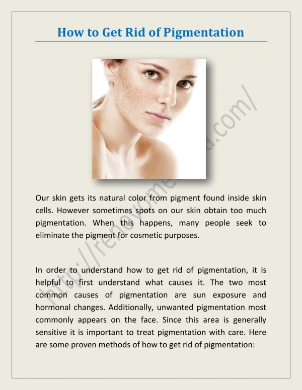
Disorders of Pigmentation. Postinflammatory Pigmentary Changes. Occurs Following Dermatitis Acne Infection Injury Description Hyperpigmentation Hypopigmentation Course Resolution over a few months. Pigmentary Mosaicism. Genetic mosaicism Born with 2 genetically different cell lines

E N D
PostinflammatoryPigmentary Changes • Occurs Following • Dermatitis • Acne • Infection • Injury • Description • Hyperpigmentation • Hypopigmentation • Course • Resolution over a few months
PigmentaryMosaicism • Genetic mosaicism • Born with 2 genetically different cell lines • Timing • May be seen at birth • May appear in infancy or early childhood • Description • Whorled, fountain-like pattern of alternating hypo- and hyperpigmentation • Blaschkoid distribution • Macules without vesicular or verrucous lesions • Other problems • CNS, MSK, eye • More likely if skin findings are prominent
Vitiligo • Acquired partial to complete loss of pigmentation • Description • Well-demarcated hypopigmented and depigmentedmacules and patches • Enhance under Wood’s lamp • Absence of melanocytes • Location • Eyes, mouth, genitals, elbows, hands and feet • Can appear anywhere • Cause • Autoimmune
Ash-Leaf Spots • Timing • Congenital • Description • Well demarcated, hypopigmentedmacules • Lancinate shape • May enhance with Wood’s light • Location • Truncal • Associated with • Tuberous Sclerosis • Normal • 0.5% newborns • Nevus depigmentosus • Localized form of pigmentarymosaicism
Albinism • Timing • Congenital • Description • Hypopigmentation of the skin, eyes and hair • X-linked ocular • Skin is normal • AR • Oculocutaneous • Many variants • Complications • At risk of skin cancer • Must avoid excessive sun and use sunscreen
Albinism • Variants • Tyrosinase-negative OCA • No trace of pigment • Snow-white hair, pinkish-white skin, translucent or blue irises • Also may have nystagmus, moderate to severe strabismus and poor visual acuity • Tyrosinase-positive OCA • Look similar at birth • Develop variable amounts of pigment with increasing age • Eye color – gray to light brown • Hair color – blonde or light brown
Piebaldism • Partial albinism • AD – rare • Description • White forelock • Triangular patch of depigmentation and white hair on the frontal scalp • Circumscribed congenital leukoderma • Hypopigmented or depigmentedmacules on the face, neck, ventral trunk, flanks or extremities • May have scattered patches of normal pigmentation within
Piebaldism • Variants • Waardenburg syndrome • Lateral displacement of the inner canthi and inferior lacrimal ducts • Flattened nasal bridge • Sensorineural deafness • Wolf syndrome • AR • Neurologic deficits
Café-au-lait spots • Description • Tan macules • May be a marker for • Neurofibromatosis type 1 • Small and smooth borders • McCune-Albright • Large and segmental with jagged borders • Large or >4
Question 10 An 8-year-old boy presents with a 2-week history of an enlarging, tender lump on the scalp. The only notable finding on physical examination are alopecia overlying a boggy mass on the scalp and posterior cervical lymphadenopathy. Of the following, the MOST appropriate treatment is • Cefazolin orally • Griseofulvin orally • Incision and drainage • Ketoconazole topically • Mupirocin topically
Alopecias • Alopecia Telogen Effluvium • Partial, temporary • 3 months after an emotional or physical stress • <50% loss • Alopecia Anagen Effluvium • Sudden loss of growing hairs • 80% • Tapering of the hair shaft and loss of adhesion to the follicle • Chemotherapy
Alopecias • Alopecia Areata • Description • Round or oval patches of hair loss • Absence of inflammation and scaling • Short (3-6mm) easily removable hairs at the margins • Location • Scalp, eyebrows, lashes or body • Severity • May be diffuse or generalized • Cause • Autoimmune • Other findings • Scotch-plaid pitting of the nails • Course • Difficult to predict • Treat any comorbid conditions
Alopecias • TrichorrhexisNodosa • Timing • At any age • Description • Hair shaft breakage • Brittle, short hairs • Frayed edges by microscopy • Cause • Damage to the outer cortex of the hair shaft • Trauma, chemicals • Course • Resolution with removal of insult
Alopecias • Friction alopecia • Infants • Self-limited • Prevented/treated with tummy-time • If severe or long standing consider neglect • Traction alopecia • Maintaining a tight pull on the hair shafts • Ponytails, pigtails, braids, cornrows • Course • Shaft fractures and follicular damage • Can lead to permanent scarring
Alopecias • Trichotillosis • AKA trichotillomania • Timing • School-age children and adolescents • Description • Bizarre patterns of hair loss • Usually broad linear bands on vertex or sides of scalp • Side opposite of dominant hand • Short, broken-off hairs with stubs of different lengths • Never completely bald • Associations • Situational stress • Sometimes psychiatric
Aplasia Cutis Congenita • AR • Timing • Congenital • Etiology • Absence of or failure of formation of a localized area of scalp or skin • Dermis and epidermis • Some involve subq tissue and rarely calvarial defects • Location • Single lesion on vertex of scalp • Rarely multiple or involving trunk or extremities
Aplasia Cutis Congenita • Description • Birth • Sharply circumscribed open and weeping ulceration that may be covered by a thin membrane or crust • Healed in several weeks to months • Usually smooth atrophic scar • Look for hair collar sign • Neural tube defects • Associations • Limb defects, genetic anomalies • Treatment • Saline compresses • Topical antibiotics • Sterile dressings
TineaCapitis • Most common organism • Trichophytontonsurans • Does not fluoresce • >95% • Microsporumcanis • Fluoresces bluish-green • 5% • Description is variable • Mild erythema and scaling with partial alopecia • Widespread breakage of the scalp • “salt-and-pepper” • More erythema, edema and pustules • Crusting • Heaped up scale with small pustules
TineaCapitis • Description is variable • Kerion • Intense inflammation with raised, tender, boggy plaques or masses studded with pustules • KOH mount shows infected hairs • Associated occipital, postauricular and posterior cervical adenopathy • Treatment • Oral antifungal agents • 6 weeks to 4 months • Selenium sulfide shampoo • Steroids • Severe inflammation • Course • May lead to scarring and permanent hair loss if untreated • Prevention • Do not share!
Congenital and Genetic Disorders • Monilethrix • AD • Developmental hair defect • Timing • 2-3 month of age • Description • Brittle, beaded hair • Periodic narrowing of the hair shaft • Location • Scalp most severely affected • Course • Permanent • Appearance may improve with age
Congenital and Genetic Disorders • Pili torti • Hair shaft is twisted on its own axis • Timing • Appears with first terminal hair growth of infancy • Location • Localized or generalized • Associations • Menkes kinky hair syndrome • Defect of copper absorption • CNS, CV, Skeletal systems
Disorders Affecting Nails • Paronychia • Acute • Red, swollen, tender nail fold • Bacterial invasion • Staph or strep • Chronic • One or several nails • Chronic dermatitis or frequent exposure to water • Nail dystrophy • Candida species • Treat with topical antimycotics
Disorders Affecting Nails • Onychomycosis • Fungal infection • Uncommon before puberty • Dystrophic nails • More commonly a result of trauma • Or underlying dermatosis
Disorders Affecting Nails • Subungual hemorrhage • Trauma • Crush injuries • Turf toe • Jamming toe into the end of a shoe suddenly • Description • Purplish-brown pigment • Treatment • Evacuation if painful