Download
1 / 2
30 likes | 197 Vues
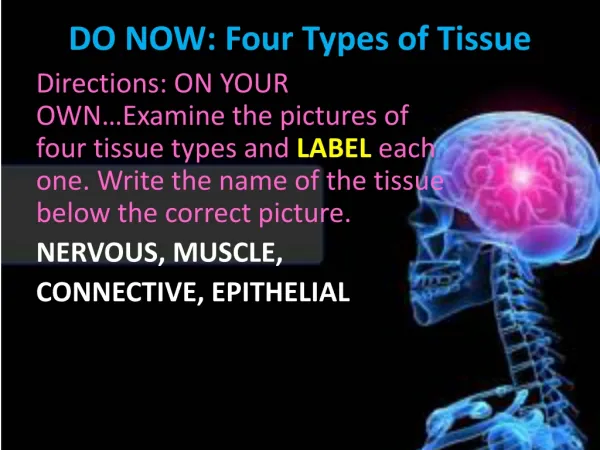
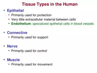
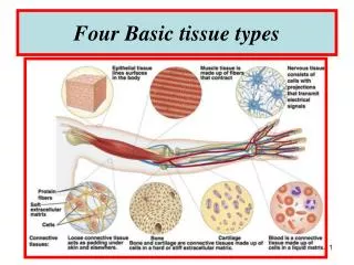
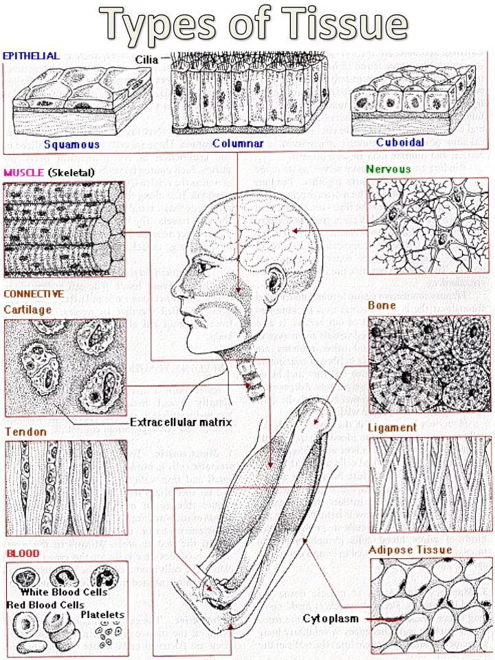
Types of Tissue . 1. Epithelial Epithelial tissue is made of closely-packed cells arranged in flat sheets. Epithelia form the surface of the skin, line the various cavities and tubes of the body, and cover the internal organs. Subsets of Epithelia

E N D
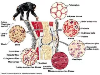
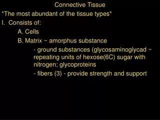
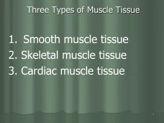
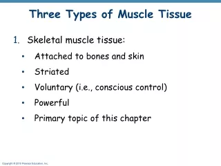
1. Epithelial • Epithelial tissue is made of closely-packed cells arranged in flat sheets. Epithelia form the surface of the skin, line the various cavities and tubes of the body, and cover the internal organs. • Subsets of Epithelia • Epithelia that form the interface between the internal and external environments. • Skin as well as the lining of the mouth and nasal cavity. These are derived from ectoderm. • Inner lining of the GI tract, lungs, urinary bladder, exocrine glands, vagina and more. These are derived from endoderm. • The apical surface of these epithelial cells is exposed to the "external environment", the lumen of the organ or the air. • Mesothelia. These are derived from mesoderm. • pleura — the outer covering of the lungs and the inner lining of the thoracic (chest) cavity. • peritoneum — the outer covering of all the abdominal organs and the inner lining of the abdominal cavity. • pericardium — the outer lining of the heart. • Endothelia. The inner lining of the heart, all blood and lymphatic vessels — derived from mesoderm. • The basolateral surface of all epithelia is exposed to the internal environment (ECF). The entire sheet of epithelial cells is attached to a layer of extracellular matrix that is called the basement membrane or, better (because it is not a membrane in the biological sense), the basal lamina. • The function of epithelia always reflects the fact that they are boundaries between masses of cells and a cavity or space. Some examples: • The epithelium of the skin protects the underlying tissues from mechanical damage, ultraviolet light, dehydration, invasion by bacteria • The columnar epithelium of the intestine secretes digestive enzymes into the intestine; absorbs the products of digestion from it. • An epithelium also lines our air passages and the alveoli of the lungs. It secretes mucus which keeps it from drying out and traps inhaled dust particles. Most of its cells have cilia on their apical surface that propel the mucus with its load of foreign matter back up to the throat. • 2. Muscle • Three kinds of muscle are found in vertebrates: Skeletal muscle is made of long fibers whose contraction provides the force of locomotion and other voluntary body movements. Smooth muscle lines the walls of the hollow structures of the body, such as the intestine, urinary bladder, uterus, and blood vessels. Its contraction, which is involuntary, reduces the size of these hollow organs. The heart is made of cardiac muscle. • 3. Connective • The cells of connective tissue are embedded in a great amount of extracellular material. This matrix is secreted by the cells. It consists of protein fibers embedded in an amorphous mixture of protein-polysaccharide ("proteoglycan") molecules. Supporting connective tissue:Gives strength, support, and protection to the soft parts of the body. cartilage. Example: the outer ear bone. The matrix of bone contains collagen fibers and mineral deposits. The most abundant mineral is calcium phosphate, although magnesium, carbonate, and fluoride ions are also present. Dense connective tissue: Often called fibrous connective tissue. Tendons connect muscle to bone. The matrix is principally Type I collagen, and the fibers are all oriented parallel to each other. Tendons are strong but not elastic. Ligaments attach one bone to another. They contain both collagen and also the protein elastin. Elastin permits ligaments to be stretched. Loose connective tissue: It is distributed throughout the body. It serves as a packing and binding material for most of our organs. Sheets of loose connective tissue that bind muscles and other structures together are called fascia. Collagen, elastin, and other proteins are found in the matrix of loose connective tissue. Both dense and loose connective tissue are derived from cells called fibroblasts, which secrete the extracellular matrix. Adipose tissue:Adipose tissue is "fat". There are two kinds found in mammals: white adipose tissue (WAT) in which the cells, called adipocytes, have become almost filled with oil. The oil is confined within a single membrane-enclosed droplet. Virtually all of the "fat" in adult humans is white adipose tissue. brown adipose tissue (BAT) in which the adipocytes contain many small droplets of oil as well as many mitochondria. White adipose tissue and brown adipose tissue differ in function as well as cellular structure. New adipocytes in white adipose tissue are formed throughout life from a pool of precursor cells. These are needed to replace those that die (after an average life span of 10 years). Whether the total number of these adipocytes increases in humans becoming fatter as adults is still uncertain. If not, why do so many of us get fatter as we age? Because of the increased size of individual adipocytes as they become filled with oil. The adipocytes of white adipose tissue secrete several hormones, including leptin and adiponectin. • Blood: The bone marrow is the source of all the cells of the blood. These include: red blood cells (RBCs or erythrocytes) five kinds of white blood cells (WBCs or leukocytes) and platelets (or thrombocytes). • 4. Nerve • Nerve tissue is composed of nerve cells called neurons and glial cells. • Neurons:Neurons are specialized for the conduction of nerve impulses. A typical neuron consists of a cell body which contains the nucleus; • a number of short fibers — dendrites — extending from the cell body a single long fiber, the axon. • The nerve impulse is conducted along the axon. The tips of axons meet: other neurons at junctions called synapses and muscles (called neuromuscular junctions) glands. • Glial cells surround neurons. Once thought to be simply support for neurons (glia = glue), they turn out to serve several important functions. There are three types: • Schwann cells. These produce the myelin sheath that surrounds many axons in the peripheral nervous system. • Oligodendrocytes. These produce the myelin sheath that surrounds many axons in the central nervous system (brain and spinal cord). • Astrocytes. These — often star-shaped — cells are clustered around synapses and the nodes of Ranvier where they perform a variety of functions: stimulating the formation of new synapses; modulating the activity of neurons; supplying neurons with materials (e.g. glucose and lactate) as well as some signaling molecules; regulating the flow of blood to their region of the brain. It is primarily the metabolic activity of astrocytes that is being measured in brain imaging by positron-emission tomography (PET) and functional magnetic resonance imaging (fMRI). • In addition, the central nervous system contains many microglia — mobile cells (macrophages) that respond to damage (e.g., from an infection) by engulfing cell debris secreting inflammatory cytokines like tumor necrosis factor (TNF-α) and interleukin-1 (IL-1) Microglia are also active in the healthy brain, at least in young mice, where they engulf synapses thus reducing the number of synapses in the developing brain.