Download
1 / 40
410 likes | 804 Vues
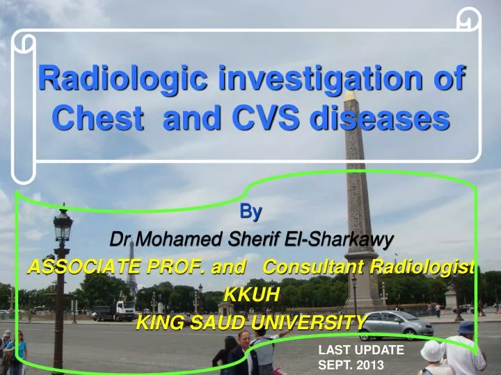
Radiologic investigation of Chest and CVS diseases. By Dr Mohamed Sherif El-Sharkawy ASSOCIATE PROF. and Consultant Radiologist KKUH KING SAUD UNIVERSITY. LAST UPDATE SEPT. 2013. What do we mean by chest . We mean study of thoracic cage contents. LUNGS . BASIC CHEST EXAMS.

E N D
Radiologic investigation of Chest and CVS diseases By Dr Mohamed Sherif El-Sharkawy ASSOCIATE PROF. and Consultant Radiologist KKUH KING SAUD UNIVERSITY LAST UPDATE SEPT. 2013
What do we mean by chest We mean study of thoracic cage contents
BASIC CHEST EXAMS PLAIN FILM=CHEST X-RAY(CXR) CT HRCT ANGIOGRAMS
Imaging Modalities for chest and CVS examinations 1-Plain films 2-COMPUTED TOMOGRAPHY CT LUNGS AND MEDIASTINUM CT- angiography (CTA) High resolution CT of the chest (HRCT) 3-Angiography 4- MRI
PA vs. AP FALSE ELARGEMENT
Inspiration • This greatly helps the radiologist to determine if there are intrapulmonary abnormalities. • The diaphragm should be found at about the level of the 8th - 10th posterior rib or 5th - 6th anterior rib on good inspiration.
Rotation • The technologists are usually very careful to x-ray the patient flat against the cassette. If there is rotation of the patient, the • Mediastinum may look very unusual. • One can access patient rotation by observing the clavicular heads and determining whether they are equal distance from the spinous process of the thoracic vertebral bodies.
In this rotated film skin folds canbe mistaken for a tension pneumothorax (blue arrows). Notice the skewed positioning of the heads of the clavicles (red arrows) and the spinous processes.
Anatomy on Normal Chest X-Ray Heart borders and chambers of the heart on PA and lateral views.
Diagram of lungs showing lobes. The right lung has threelobes, upper, middle and lower. These are separated by the oblique and horizontal fissures. The left lung has two lobes, upper and lower separated by the oblique fissure.
(1) Horizontal fissure(2) Right oblique fissure, (3) Left oblique fissure. Figure 2.4b(1) Horizontal fissure (2) Right oblique fissure (3) Right upper lobe(4) Right middle lobe (5) Right lower lobe. Figure 2.4c (1) Leftoblique fissure (2) Left upper lobe (3) Left lower lobe. Why these lines are important LEFT LUNG RIGHT LUNG
CARDIAC Valves This patient had a malfunctioning mitral valve (between left atrium and left ventricle) and aortic valve (between left ventricle and aorta) and prosthetic valves were inserted (better seen on lateral) Frontal CXR LAT CXR • Key: • Suture material used for repair of vertical incision thru sternum (median sternotomy) • Aortic valve prosthesis • Mitral valve prosthesis • Left hemi diaphragm • Right hemi diaphragm 1 1 2 2 3 3 5 4 4 5
MITRAL VALVE REPLACEMENT LLL COLLAPSE KKUH
ROUTINE CXR BREAST SHADOW
Frontal Chest X-Ray SILHOUETTE See Section on the Silhouette Sign
Frontal Chest X-Ray MEDIASTINUM
The Aortic arch/great vessels “Man’s Anatomy by Tobias & Arnold
Aortic aneurysm Aortic knob/knuckle
HRCT uses very thin slices (1mm) to achieve better spatial resolution & precision. HRCT is indicated after normal CXR in a symptomatic patient - the setting of high clinical suspicion of disease. Advantages High sensitivity for adenopathy, infiltrates, and architectural distortion. HRCT can identify areas of reversible vs. irreversible lung damage. High Resolution CT Scan
Tracheobronchial Tree Normal lung at level inferior pulmonary veins L inferior pulmonary vein R inferior pulmonary vein Lower lobe bronchi Normal Lung Anatomy
Anatomy on Normal Chest X-Ray CXR-PA 1 2 2 4 13 • Key: • Right 1st rib • Right 2nd rib • Scapula • Trachea • Carina • Bronchus seen end on • Bilateral hila • Branch of right main descending pulmonary artery • Right minor (horizontal fissure) • Right hemi diaphragm • Left hemi diaphragm • Gastric air bubble • Left clavicle 3 5 6 7 7 8 9 10 11 12
Frontal Chest X-Ray Intrapulmonary nodule: hamartoma Nodule or right nipple ?
Remember It’s a chest x-ray, not a lung x-ray.