Download
1 / 12
170 likes | 454 Vues
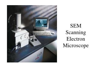
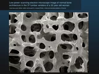
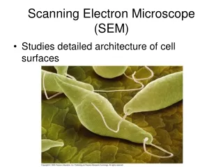
Scanning Electron Microscope . Jamie Goings. Theory. Conventional microscopes use light and glass lenses SEM uses electrons and magnetic lenses to create magnification Electron beam ‘traces’ over object, interacting with surface and dislodges surface electrons

E N D
Scanning Electron Microscope Jamie Goings
Theory • Conventional microscopes use light and glass lenses • SEM uses electrons and magnetic lenses to create magnification • Electron beam ‘traces’ over object, interacting with surface and dislodges surface electrons • Detector collects electrons, and registers different levels of brightness • Scanned onto monitor dot by dot, row by row
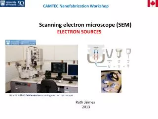
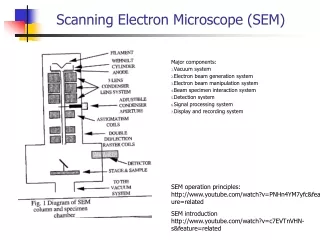
Instrumentation – Electron Gun 1. Thermionic Guns – most common voltage is applied that heats filament (usually tungsten) 2. Field Emission Guns Creates strong electrical field to pull e away from atoms Anode attracts e causing them to accelerate down into a beam
II. Lenses Magnetic lenses direct electron beam into wanted path. III. Scanning Coils Additional magnetic field use voltage to move beam back and forth across sample. Can adjust magnification here by adjusting scan area IV. Sample Chamber must be sturdy and isolated from vibration adjustable position vacuum chamber – keeps e beam clear of air particles and sample free of dust
V. Detectors 1. Secondary Electron Detector has a 300V positive charged metal cage to attract e collects e dislodged from surface number of e collected per ‘dot scan’ determined brightness of spot creates image 2. Energy Dispersive X-Ray Detector (EDX) elemental analysis analyzes x-rays emitted from specimen detects number of x-rays vs their energy- energy of x-ray is specific to element it was emitted from
Sample / Sample Prep Sample must be conductive must be able to withstand vacuum – no liquids 15/15 mm older machines 200/200 mm modern Sample Prep clean dust or debris sputter coating biological samples are dehydrated and dried
Benefits / Limitations Benefits High depth of field High resolution – high magnification Can adjust focus, contrast, brightness Computer controls 3D image With EDX is both qualitative and quantitative Limitations Generates radiation Needs to be clean!!!
Work Cited: Oatley, C. W. The Scanning Electron Microscope Gabriel, Biological Scanning Electron Miscroscopy Springer-Verlag, Methods of Preparation for Electron Microscopy http://www.purdue.edu/rem/rs/graphics/sem2.gif http://www.herguth.com/technical/sem.pdf http://static.howstuffworks.com/gif/scanning-elecron-microscope-illustration.jpg (image)