Download
1 / 13
130 likes | 265 Vues
PREPARATION OF BIOTIN-GLUTATHIONE COATED QUANTUM DOTS. Libor Janů Supervisor: doc. Ing. René Kizek, PhD. Department of Chemistry and Biochemistry, Faculty of agronomy, Mendel University. Introduction. Semiconductor nanocrystals (2-10 nm)

E N D
PREPARATION OF BIOTIN-GLUTATHIONE COATED QUANTUM DOTS Libor Janů Supervisor: doc. Ing. René Kizek, PhD. Department of Chemistry and Biochemistry, Faculty of agronomy, Mendel University
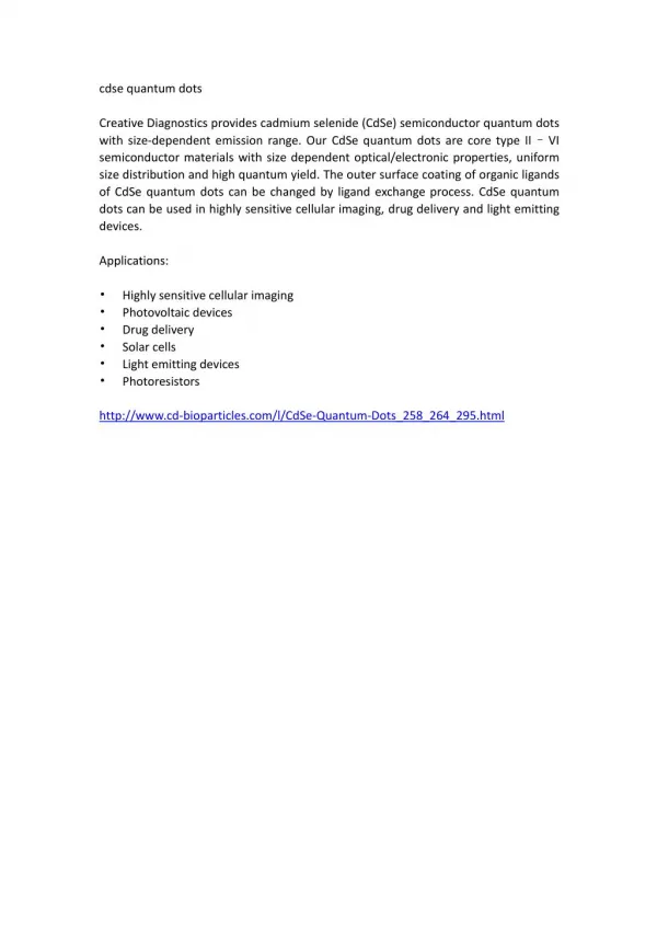
Introduction • Semiconductor nanocrystals (2-10 nm) • Fluorescent labeles in a variety of biological investigations (biological active compounds, Hg2+,…) • Better spectroscopic properties and longer lifetime in comparison to classical molecular dyes
Introduction • Solution phase synthesis of quantum dots • Usually CdTe or CdSe QD • Low quantum yield and toxic properties of naked QD • Modifications: TOPO (trioctylphosphine oxide) coating – biocompatible, better spectroscopic properties but hydrophobic • Solubility improving: ligand exchange (mercaptoacetic acid or DHLA), silanization, amphiphilic copolymers, … • Long reaction times, multistep process, high temperatures, inert atmosphere.
Introduction Fig.1: Quantum dots modifications. Source: Min Zhou, Indraneel Ghosh, Current Trends in Peptide Science, Quantum Dots and Peptides: A Bright Future Together, PeptideScience Volume 88 / Number 3, 327 Thiol chelation Phospholipids Silanization Copolymer Ligand exchange Hydrophobic interaction
Material and methods The aim of the work: • Biotinylated quantum dots • Glutathione as the coating agent providing solubility in water • One step synthesis in water Synthesis of biotin-glutathione • Solid phase peptide synthesis on Pioneer Peptide Synthesiser • Biotin-glutathione was synthesized using peptide bonding ofthe biotin carboxy group and amino group of the gamma-glutamic acid GSH QD Biotin + avidin-biomolecule
Material and methods Synthesis of biotin-glutathione quantum dots • Reaction CdCl2 + Na2TeO3 in the presence of the biotin-glutathione • Mercapto group of glutathione thiolates the quantum dot surface • Carboxilic acid provides solubility in water • Na2TeO3 is air stable avoiding the need of inert atmosphere during synthesis • Reaction conditions: 95°C, synthesis time 2,5 hour Reaction: 4TeO32- +3BH4- → 4Te2- +3BO2- +6H2O CdCl2 + Te2- + MPA → Cd- (MPA)xTey+2Cl- Cd- MPAxTey → CdTe Junling Duan et.al.: One-Pot Synthesis of Highly Luminescent CdTe Quantum Dots by Microwave Irradiation Reduction and Their Hg2+-Sensitive Properties. Nano Res (2009) 2: 61 68
Results B800 B-GSH Fig. 2: MALDI-TOF MS of biotin-glutathione. Peaks with the diferent mass are HCCA matrix and Na+ adducts. Purity of crude product ≥ 80%, purity of pure peptide ≥ 98%. (Ryvolova et. al.:Biotin-modified glutathione as a functionalized coating for bioconjugation of CdTe-based quantum dots. Electrophoresis 2011, 32, 1619–1622) B-GSH Intensity (a.u.) [M-2H+Na]- HCCA matrix HCCA matrix HCCA matrix m/z
Results B-GSH Absorbance 214 nm (AU) Fig.3: Electropherogram of crude quantum dot-biotin-glutathione . UV detection at 214 nm. (Ryvolova et. al.:Biotin-modified glutathione as a functionalized coating for bioconjugation of CdTe-based quantum dots. Electrophoresis 2011, 32, 1619–1622) GSH B-GSH – QD Migration Time (min)
Results Fig.4: Electropherogram of crude quantum dot-biotin-glutathione. LIF detection (488 nm/530 nm); inset: B-GHS-QDs under ambient light (left), quantum dot-biotin-glutathione under UV light (right). (Ryvolova et. al.:Biotin-modified glutathione as a functionalized coating for bioconjugation of CdTe-based quantum dots. Electrophoresis 2011, 32, 1619–1622) B-GSH – QD Fluorescence (a.u.) Migration Time (min)
Results Fig.5: Electropherogram of the mixture of the B-GSH-QDs and avidin solution; LIF detection (488 nm/530 nm). (Ryvolova et. al.:Biotin-modified glutathione as a functionalized coating for bioconjugation of CdTe-based quantum dots. Electrophoresis 2011, 32, 1619–1622) B-GSH – QD Avidin-B-GSH – QD
Conclusion • Biotinylated quantum dots as a probe with specific afinity to avidin and streptavidin • No need the multistep and time consuming synthesis in organic solvents. • Next synthesis efficiencyenhancement possibilities: microvawe synthesis – homogenous heating of the sample,smaller surface defects, shorter reaction times.
Acknowledgements • doc. Ing. René Kizek, PhD. • Ing. Markéta Ryvolová, PhD. • Ing. Jana Chomoucká, PhD. • Mgr. Natalia Cernei • Ing. Pavlína Šobrová • Mgr. Ondřej Zítka • This study was supported fromIGA AF Mendelu,IP 13/2011.