Download
1 / 38
410 likes | 764 Vues
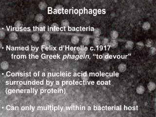
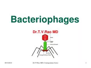
Bacteriophages. Self assembly and Applications. Bacteriophages: Definition & History. Bacteriophages are viruses that can infect and destroy bacteria. They have been referred to as bacterial parasites, with each phage type depending on a single strain of bacteria to act as host. .

E N D
Bacteriophages Self assembly and Applications
Bacteriophages: Definition & History • Bacteriophages are viruses that can infect and destroy bacteria. • They have been referred to as bacterial parasites, with each phage type depending on a single strain of bacteria to act as host.
Bacteriophages: Classification • Based on two major criteria: • phage morphology (electron microscopy) • nucleic acid properties
Bacteriophages: Classification • At present, over 5000 bacteriophages have been studied by electron microscopy and can be divided into 13 virus families.
BACTERIAL CELL INFECTION BY VIRUS * Virus binds to receptor and ejects genome *Viral particlestays outside cell! Only its genome enters*Virion leaves via lysis of cell
13 Bacteriophage families Double stranded DNA, Non-enveloped Double stranded DNA, Enveloped SIRV 1, 2 P2 Rudiviridae Myoviridae Plasmaviridae T2 Fuselloviridae SSV1 TTV1 Tectiviridae λ PRD1 Siphoviridae Lipothrixviridae PM2 P22 Corticoviridae Podoviridae Single stranded RNA Double stranded RNA Single-stranded DNA M13 & fd Inoviridae MS2 phi666 Leviviridae ΦX174 Microviridae Cystoviridae
Bacteriophages: Virulence Factors Carried On Phage • Temperate phage can go through one of two life cycles upon entering a host cell. • Lytic: Is when growth results in lysis of the host and release of progeny phage. • Lysogenic: Is when growth results in integration of the phage DNA into the host chromosome or stable replication as a plasmid. Most of the gene products of the lysogenic phage remains dormant until it is induced to enter the lytic cycle.
Bacteriophages: Lysogenic Conversion • Some lysogenic phage carry genes that can enhance the virulence of the bacterial host. • For example, some phage carry genes that encode toxins. • These genes, once integrated into the bacterial chromosome, can cause the once harmless bacteria to release potent toxins that can cause disease.
Bacteriophages: Lysogenic Conversion Examples of Virulence Factors Carried by Phage
Bacteriophages • The filamentous Phage f: • A: AFM image • B: Schematic representation • 1: initiation of assembly • 2,3: elongation • 4: termination
Bacteriophages • Used for cloning foreign genes among other applications • Proteins and peptides are fused to the Capsid(surface) of the phage • The combination of the phage and peptide is known as a Fusion Protein
Bacteriophages • Different sets of genes are inserted into the genomes of multiple phages • These separate phages will only display one protein, peptide, or antibody • Collections of these phages can comprise Libraries • These Libraries are exposed to selected targets and only some phages will interact with targets
Bacteriophages • 3 types of common phages used in phage display are the M13, F1 , FD • Virions take up a small amount of area • Through using multiple Virions polypeptide libraries can be created, and each phage displays a random peptide
Bacteriophages • By taking gene segment of antigens of antibodies and fusing them to the protein coat of phages, these phages will now express the anti-body in a fusion protein • Phage Display Libraries of antigens can be created to create anti-body phage display libraries
Bacteriophages • Polypeptides of interest can be screened using selection techniques • The target protein/peptide can be immobilized using magnetic beads • With the advance of DNA sequence recognition these selected sequences can be identified easily
Bacteriophages • Phage display
Bacteriophages • Once these Phages are isolated and recovered they can be used to infect bacteria which will create a particle similar to a monoclonal antibody
Bacteriophages A: wt; B-F: types of pIII displays; G: pVII or pIX display H: mosaic pVIII display, I:uniform pVIII display
Bacteriophages • Morphology of the T series of Phages
Bacteriophages The Phages HK97 and HK022 do have a very prominent friend the bacteriophage λ
Properties of Filamentous Viruses fd Pf1 Pf3 PH75 Symmetry class I (C5S2)a II (C1S5.4)b II (C1S5.4)b II (C1S5.4)c Length (nm) 880 1900 680 910 Ext. diam. (nm) 6.5 6.5 6.5 6.5 No. subunitsd 2750 7400 25002700 No. nucleotides 6408 7420 5800 6500 Nucl./subunit 2.4 1.0 2.4 2.4 Wt-% protein 87 94 86 87 a10 subunits per 32 Å helical repeat. Marvin et al. (2006) J. Mol. Biol. 355, 294-309. b27 subunits per 75 Å helical repeat. Welsh et al. (2000) Acta Cryst. D56, 137-150; Welsh et al. (1998) J. Mol. Biol. 283, 155-177. cPederson et al. (2001) J. Mol. Biol. 309, 401-421. dSequences: fd: AEGDDPAKAA FDSLQASATEYIGYAWAMVV VIVGATIGIK LFKKFTSKAS50 (5.24 kDa; pI = 6.3) Pf1: GVIDTSAVES AITDGQGDMK AIGGYIVGAL VILAVAGLIY SMLRKA46 (4.61 kDa; pI = 4.7) Pf3: MQSVITDVTG QLTAVQADIT TIGGAIIVLA AVVLGIRWIK AQFF44 (4.63 kDa; pI = 5.7) PH75: MDFNPSEVAS QVTNYIQAIA AAGVGVLALA IGLSAAWKYA KRFLKG46 (4.81 kDa; pI = 9.4) 060611
fd Architecture ssDNA core 65 Å 1/100th virion length Filament of 6.5 x 880 nm (PL = 2 μm) Coat of layered -helical subunits Arranged with 5-fold rotational symmetry Right-hand slew on capsid surface ssDNA packaged within (conformation?) Caspar & Makowski (1981) J. Mol. Biol. 145, 611-617. Day et al. (1988) Ann. Rev. Biophys. 17, 509-539. Marvin et al. (1994) J. Mol. Biol. 235, 260-286. fd (6.5 x 880 nm) 060611
Molecular Models of the fd Capsid solid state NMR fiber X-ray diffraction Zeri et al. (2003) PNAS 100, 6458-6463. Marvin et al. (2006) J. Mol. Biol. 355, 294-309. 060611