Download
1 / 2
60 likes | 289 Vues
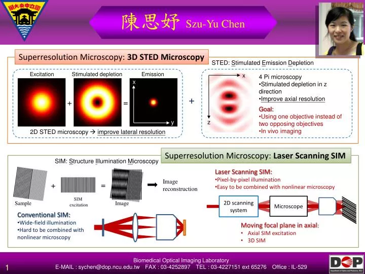
陳思妤 Szu-Yu Chen. Superresolution Microscopy: 3D STED Microscopy. STED: S timulated E mission D epletion. Excitation. Stimulated depletion. Emission. x. 4 Pi microscopy Stimulated depletion in z direction Improve axial resolution Goal:

E N D
陳思妤Szu-Yu Chen Superresolution Microscopy: 3D STED Microscopy STED: Stimulated Emission Depletion Excitation Stimulated depletion Emission x • 4 Pi microscopy • Stimulated depletion in z direction • Improve axial resolution • Goal: • Using one objective instead of two opposing objectives • In vivo imaging x + + = z y 2D STED microscopy improve lateral resolution Superresolution Microscopy: Laser Scanning SIM SIM: Structure Illumination Microscopy • Laser Scanning SIM: • Pixel-by-pixel illumination • Easy to be combined with nonlinear microscopy Image reconstruction = + Sample Image 2D scanning system Microscope • Conventional SIM: • Wide-field illumination • Hard to be combined with nonlinear microscopy • Moving focal plane in axial: • Axial SIM excitation • 3D SIM Biomedical Optical Imaging Laboratory E-MAIL : sychen@dop.ncu.edu.twFAX : 03-4252897TEL : 03-4227151 ext 65276Office : IL-529
陳思妤Szu-Yu Chen 4D Hyperspectral microscopy Image: Intensity recorded pixel-by-pixel within certain wavelength range Spectrum: Intensity recorded for a single spot at multiple wavelength Hyperspectral image: Intensity recorded at multiple wavelength pixel-by-pixel Image (Y) Detector Spectrometer 2D detector array Wavelength (nm) Image (X) + • Biomedical information: • Morphological (intensity) and molecular (spectral) information • Biomedical applications: • Molecular imaging • Disease diagnosis and treatment Conventional imaging Spectroscopy Hyperspectral imaging 4 Ds: 2 dimensions of image, wavelength, and time Biomedical Optical Imaging Laboratory E-MAIL : sychen@dop.ncu.edu.twFAX : 03-4252897TEL : 03-4227151 ext 65276Office : IL-529