Download
1 / 96
970 likes | 1.2k Vues
Animals and Animal Diversity. The Nitty-gritty!. Note:. There is no red on this powerpoint, all non-essentials were deleted from the notes. Just imagine that everything is in red!. Ch 32?. Basic Characteristics. Multicellular Heterotrophic Mobile Eukaryotic Lack cell walls

E N D
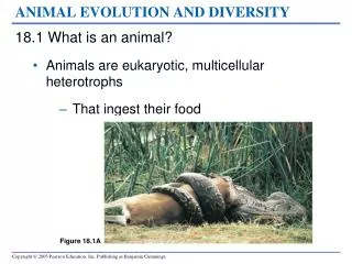
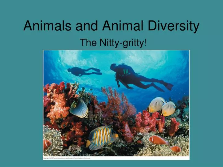
Animals and Animal Diversity The Nitty-gritty!
Note: • There is no red on this powerpoint, all non-essentials were deleted from the notes. • Just imagine that everything is in red!
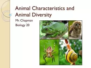
Ch 32? Basic Characteristics • Multicellular • Heterotrophic • Mobile • Eukaryotic • Lack cell walls • Bodies are held together by structural proteins like collagen • Nervous and muscular tissue unique to animal kingdom
Reproduction and Development • Most reproduce sexually, with the diploid stage usually dominating the life cycle • After a sperm fertilizes an egg, the zygote undergoes rapid cell division called cleavage • Cleavage leads to formation of a blastula • The blastula undergoes gastrulation, forming a gastrula with different layers of embryonic tissues Video: Sea Urchin Embryonic Development
Fig. 32-2-3 Blastocoel Endoderm Cleavage Cleavage Blastula Ectoderm Archenteron Eight-cell stage Zygote Gastrulation Gastrula Blastocoel Blastopore Cross section of blastula
Many animals have at least one larval stage (sexually immature morphology that is different from the adult), which eventually undergoes metamorphosis • All animals, and only animals, have Hox genes that regulate the development of body form
Paleozoic Era (542–251 Million Years Ago) – The rise of the animal kingdom • The Cambrian explosion (535 to 525 million years ago) marks the earliest fossil appearance of many major groups of living animals • There are several hypotheses regarding the cause of the Cambrian explosion • New predator-prey relationships • A rise in atmospheric oxygen • The evolution of the Hox gene complex
Concept 32.3: Animals can be characterized by “body plans” • Zoologists sometimes categorize animals according to a body plan, a set of morphological and developmental traits
Symmetry Radial • Animals can be categorized according to the symmetry of their bodies, or lack of it • Some animals have radial symmetry, while others show bilateral symmetry. Bilateral
Two-sided symmetry is called bilateral symmetry • Bilaterally symmetrical animals have: • A dorsal (top) side and a ventral (bottom) side • A right and left side • Anterior (head) and posterior (tail) ends • Cephalization, the development of a head
Tissues • Animal body plans also vary according to the organization of the animal’s tissues • Tissues are collections of specialized cells isolated from other tissues by membranous layers • During development, three germ layersgive rise to the tissues and organs of the animal embryo
Ectoderm is the germ layer covering the embryo’s surface • Endoderm is the innermost germ layer and lines the developing digestive tube, called the archenteron • Diploblastic animals have ectoderm and endoderm • Triploblastic animals also have an intervening mesoderm layer; these include all bilaterians
Body Cavities • Most triploblastic animals possess a body cavity • A true body cavity is called a coelom and is derived from mesoderm • Coelomates are animals that possess a true coelom
Fig. 32-8a Coelom Body covering (from ectoderm) Tissue layer lining coelom and suspending internal organs (from mesoderm) Digestive tract (from endoderm) (a) Coelomate
A pseudocoelom is a body cavity derived from the mesoderm and endoderm • Triploblastic animals that possess a pseudocoelom are called pseudocoelomates
Fig. 32-8b Body covering (from ectoderm) Pseudocoelom Muscle layer (from mesoderm) Digestive tract (from endoderm) (b) Pseudocoelomate
Triploblastic animals that lack a body cavity are called acoelomates
Fig. 32-8c Body covering (from ectoderm) Tissue- filled region (from mesoderm) Wall of digestive cavity (from endoderm) (c) Acoelomate
Protostome and Deuterostome Development • Based on early development, many animals can be categorized as having protostome development or deuterostome development
Cleavage • In protostome development, cleavage is spiral and determinate • In deuterostome development, cleavage is radial and indeterminate • With indeterminate cleavage, each cell in the early stages of cleavage retains the capacity to develop into a complete embryo • Indeterminate cleavage makes possible identical twins, and embryonic stem cells
Fig. 32-9 Deuterostome development (examples: echinoderm, chordates) Protostome development (examples: molluscs, annelids) (a) Cleavage Eight-cell stage Eight-cell stage Radial and indeterminate Spiral and determinate (b) Coelom formation Key Coelom Ectoderm Mesoderm Archenteron Endoderm Coelom Blastopore Mesoderm Mesoderm Blastopore Solid masses of mesoderm split and form coelom. Folds of archenteron form coelom. (c) Fate of the blastopore Anus Mouth Digestive tube Mouth Anus Mouth develops from blastopore. Anus develops from blastopore.
Fig. 32-9a Deuterostome development (examples: echinoderms, chordates) Protostome development (examples: molluscs, annelids) (a) Cleavage Eight-cell stage Eight-cell stage Spiral and determinate Radial and indeterminate
Coelom Formation • In protostome development, the splitting of solid masses of mesoderm forms the coelom • In deuterostome development, the mesoderm buds from the wall of the archenteron to form the coelom
Fig. 32-9b Protostome development (examples: molluscs, annelids) Deuterostome development (examples: echinoderms, chordates) (b) Coelom formation Coelom Key Ectoderm Archenteron Mesoderm Endoderm Coelom Blastopore Mesoderm Blastopore Mesoderm Solid masses of mesoderm split and form coelom. Folds of archenteron form coelom.
Fate of the Blastopore • The blastopore forms during gastrulation and connects the archenteron to the exterior of the gastrula • In protostome development, the blastopore becomes the mouth • In deuterostome development, the blastopore becomes the anus
Fig. 32-9c Protostome development (examples: molluscs, annelids) Deuterostome development (examples: echinoderms, chordates) (c) Fate of the blastopore Anus Mouth Key Ectoderm Digestive tube Mesoderm Endoderm Anus Mouth Mouth develops from blastopore. Anus develops from blastopore.
Modeling Time • Let’s go back to the lab. • Take a sheet of paper with you • Pick up a direction sheet • Get 2 colors of dough
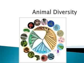
Invertebrates Those without backbones – make up about 95% of animals
Fig. 33-2 Calcarea and Silicea ANCESTRAL PROTIST Cnidaria Lophotrochozoa Common ancestor of all animals Eumetazoa Ecdysozoa Bilateria Deuterostomia
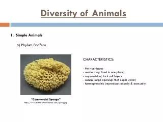
Sponges • Lack true tissues and organs • Live in water (both fresh and salt) • suspension feeders, capturing food particles suspended in the water that pass through their body • Most sponges are hermaphrodites: Each individual functions as both male and female
Fig. 33-4 Food particles in mucus Flagellum Choanocyte Collar Choanocyte Osculum Azure vase sponge (Callyspongia plicifera) Spongocoel Phagocytosis of food particles Amoebocyte Pore Spicules Epidermis Water flow Amoebocytes Mesohyl
Cnidarians • include jellies, corals, and hydras • exhibit a relatively simple diploblastic, radial body plan • body plan is a sac with a central digestive compartment, the gastrovascular cavity • A single opening functions as mouth and anus • There are two variations on the body plan: the sessile polyp and motile medusa • Carnivores that use tentacles to capture prey • Armed with enidocytes – cells that fxn in defense and capturing prey • Nematocysts – organelles that eject a stinging thread
Fig. 33-5 Mouth/anus Tentacle Polyp Medusa Gastrovascular cavity Gastrodermis Mesoglea Body stalk Epidermis Tentacle Mouth/anus
Fig. 33-6 Tentacle Cuticle of prey Thread Nematocyst “Trigger” Thread discharges Thread (coiled) Cnidocyte
Flatworms • live in marine, freshwater, and damp terrestrial habitats • acoelomates • They are flattened dorsoventrally and have a gastrovascular cavity • Gas exchange takes place across the surface
Fig. 33-10 Pharynx Gastrovascular cavity Mouth Eyespots Ganglia Ventral nerve cords
Tapeworms • Tapeworms are parasites of vertebrates and lack a digestive system • Tapeworms absorb nutrients from the host’s intestine • Fertilized eggs, produced by sexual reproduction, leave the host’s body in feces
Rotifers • Rotifers are tiny animals that inhabit fresh water, the ocean, and damp soil • Rotifers have an alimentary canal, a digestive tube with a separate mouth and anus that lies within a fluid-filled pseudocoelom • Rotifers reproduce by parthenogenesis, in which females produce offspring from unfertilized eggs • Some species are unusual in that they lack males entirely
Mollusca • Phylum Mollusca includes snails and slugs, oysters and clams, and octopuses and squids • Most molluscs are marine • Molluscs are soft-bodied animals, but most are protected by a hard shell • All molluscs have a similar body plan with three main parts: • Muscular foot • Visceral mass • Mantle • Many molluscs also have a water-filled mantle cavity, and feed using a rasplike radula
Fig. 33-15 Nephridium Heart Visceral mass Coelom Intestine Gonads Mantle Stomach Mantle cavity Mouth Shell Radula Anus Gill Radula Mouth Nerve cords Esophagus Foot
Gastropods • Most gastropods are marine, • Most have a single, spiraled shell • Slugs lack a shell or have a reduced shell • The most distinctive characteristic of gastropods is torsion, which causes the animal’s anus and mantle to end up above its head
Fig. 33-17 (a) A land snail (b) A sea slug
Fig. 33-18 Intestine Mantle cavity Stomach Anus Mouth
Bivalves • Molluscs of class Bivalvia include many species of clams, oysters, mussels, and scallops • They have a shell divided into two halves • The mantle cavity of a bivalve contains gills that are used for feeding as well as gas exchange
Fig. 33-20 Coelom Hinge area Mantle Gut Heart Adductor muscle Digestive gland Anus Mouth Excurrent siphon Shell Water flow Palp Foot Incurrent siphon Mantle cavity Gonad Gill
Cephalopods Octopus • Class Cephalopoda includes squids and octopuses, carnivores with beak-like jaws surrounded by tentacles of their modified foot • Cephalopods have a closed circulatory system, well-developed sense organs, and a complex brain Squid Chambered nautilus
Annelids • Annelids have bodies composed of a series of fused rings
Concept 33.4: Ecdysozoans are the most species-rich animal group • Ecdysozoans are covered by a tough coat called a cuticle • The cuticle is shed or molted through a process called ecdysis • The two largest phyla are nematodes and arthropods