Download
1 / 25
360 likes | 766 Vues
Lab # 7. Body Movements and Muscle Histology. Flexion, Extension and Hyperextension. Flexion : Movement that decreases the joint angle in hinge joints . Extension : Movement that straightens a joint and generally returns a body part to the zero position. Hip flexion. Lateral flexion.

E N D
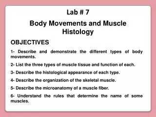
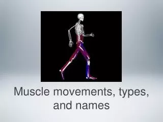
Lab # 7 Body Movements and Muscle Histology
Flexion, Extension and Hyperextension Flexion: Movement that decreases the joint angle in hinge joints Extension: Movement that straightens a joint and generally returns a body part to the zero position Hip flexion Lateral flexion Flexion Knee flexion Extension
Hyperextension: Further extension of a joint beyond the zero position. Flexion and extension occur at nearly all diarthroses, hyperextension is limited to a few joints. Hyperextension Hyperextension Extension Flexion
Abduction: Movement of a body part in the frontal plane away from the midline of the body Adduction: Movement of a body part in the frontal plane back toward the midline
Elevation: A movement that raises a body part vertically in the frontal plane Depression: A movement that lowers a body vertically part in the frontal plane Protraction: The anterior movement of a body part in the transverse (horizontal) plane Retraction: The posterior movement of a body part in the transverse (horizontal) plane
Supination: Forearm movement that turns the palm to face anteriorly or upward. The forearm is supinated in anatomical position (the radius is parallel to the ulna) Pronation: Forearm movement that turns the palm to face posteriorly or downward. The radius spins on the capitulum of the humerus. The head spins in the radial notch of ulna and the radius crosses stationary ulna like an X
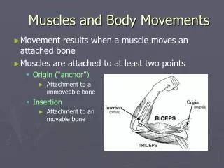
Ligaments: Bands of connective tissue that join bone to bone LIGAMENT Aponeuroses: Bands of connective tissue that attach flat muscle to another muscle or to several bones APONEUROSIS Tendons: Narrow bands of connective tissue that connect muscles to bone TENDONS
Epicraneal aponeuroses ( Galea ) Lumbar aponeuroses Abdominal aponeuroses
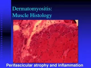
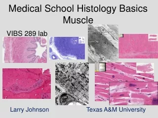
MARTINI page 133 DIFFERENT TYPES OF MUSCLE 1- Skeletal muscle 2- Cardiac muscle 3- Smooth muscle
1- Skeletal 2- Cardiac 3- Smooth MUSCLE HISTOLOGY (pages 36-39 and 77-80) Types of muscle: C e l l c h a r a c t e r i s t i c s Long, Short, Short, Cylindrical, Branched, Spindle, Striated, Striated, Non-striated, Multinuclear Single nucleus Single nucleus
Skeletal Muscle Fascicle Perimysium Endomysium Muscle fiber (cell) Epimysium
Myosatellite cell Sarcoplasm Nucleus Capillary Mitochondria Sarcolemma They are involved in the repair of damaged muscle Endomysium Myofibrils They consist of bundles of myofilaments (thin filaments and thick filaments) Axon MUSCLE FIBER (cell)
Axon of motor neuron Axon terminal Neuromuscular junction Myofilaments Sarcolemma Myofibril Motor Unit (page 79) Motor neuron It carries the nerve impulse It releases the neurotransmitter Muscle cell or fiber Nucleus (They are organized in sarcomeres)
Structure of the Skeletal Muscle Fiber Mitochondria Terminal cisterna They produce the chemical energy (ATP) for muscle contraction Sarcolemma Sarcoplasm Thin filament Myofibril Thick filament Triad Sarcoplasmic reticulum They conduct the nerve impulse from the sarcolemma to the interior of the cell It stores calcium for muscle contraction T tubules
Neuromuscular Junction and Muscle Cell or Fiber Sarcomeres Superior view Myofibrils Neuromuscular junction They release the neurotransmitter Axon terminal Nuclei Axon of the motor neuron It carries the nerve impulse Sarcolemma Sarcoplasm Endomysium
They consist of proteins called actinins, which interconnect thin filaments of adjacent sarcomeres Titin (elastic protein that attaches the thick filaments to the Z discs) Sarcomere They are the smallest functional units of the muscle fiber M line Z line Z line I band(It contains thin filaments but not thick filaments Zone of overlap H band Zone of overlap Myosin (thick filaments) Actin (thin filaments) A band M line: It consists of proteins that connect the each filament with its neighbors A band: Its length is equal to the length of the thick filaments. It contains both thin and thick filaments H band: It is a lighter region on either side of the M line, which contains only thick filaments I band Z line Zone of overlap Zone of overlap:It is the region where the thin filaments are situated between the thick filaments H band M line
A band H band I band I band Zone of overlap M line Z line Z line Zone of overlap When a skeletal muscle fiber contracts: 1- The H bands and I bands get smaller 3- The Z lines move closer together 2- The zone of overlap get larger 4- The width of the A band remain constant
Z line H zone Z line Zone of overlap Sarcomere Structure I band A band Thin filament M line Thick filament Sarcomere