Download
1 / 70
720 likes | 770 Vues
Acute Kidney Injury (AKI). Pharmacotherapy II Second Semester 2014. References. Pharmacotherapy: A Pathophysiologic Approach – Chapter 50 (Assessment) + 51 (ARF), 55 (Drug induced) Pharmacotherapy: Principles and Practice – Chapter 22

E N D
Acute Kidney Injury (AKI) Pharmacotherapy II Second Semester 2014
References • Pharmacotherapy: A Pathophysiologic Approach – Chapter 50 (Assessment) + 51 (ARF), 55 (Drug induced) • Pharmacotherapy: Principles and Practice – Chapter 22 • Applied Therapeutics: The Clinical Use of Drugs – Chapter 30 • UpToDate: http://www.uptodate.com • Assessment of kidney function: Serum creatinine; BUN; and GFR • Diagnostic approach to the patient with acute or chronic kidney disease • Urinalysis in the diagnosis of renal disease • Definition of acute kidney injury (acute renal failure) • National Kidney Foundation: http://www.kidney.org • The Renal Association. Acute kidney injury. 2011. www.renal.org/Clinical/GuidelinesSection/AcuteKidneyInjury.aspx • Kidney Disease: Improving Global Outcomes (KDIGO) Acute Kidney Injury Work Group. KDIGO Clinical Practice Guideline for Acute Kidney Injury. Kidney inter., Suppl. 2012; 2: 1–138
Introduction • Renal function: • Maintenance of body composition: • Filtration • Absorption • Secretion • Excretion of metabolic end products and foreign substances • Production and secretion of enzymes and hormones • Activation of vitamin D3/glucneogenesis/metabolism of insulin, steroids and xenobiotics
Glomerular filtration rate (GFR) • The GFR is equal to the sum of the filtration rates in all of the functioning nephrons the estimation of the GFR gives an approximate measure of the number of functioning nephrons. • The GFR is expressed as the volume of plasma filtered across the glomerulus per unit time, based on total renal blood flow (1L/min/1.73 m2)and capillary hemodynamics (plasma volume is 60% of the blood volume). • The normal value for GFR depends on age, sex, and body size, and is approximately 130 and 120 mL/min/1.73 m2 for men and women, respectively, with considerable variation even among normal individuals • A reduction in GFR implies either progression of the underlying disease or the development of a superimposed and often reversible problem, such as decreased renal perfusion due to volume depletion. • An increase in GFR, is indicative of improvement in renal function, or may imply an increase in filtration (hyperfiltration) due to hemodynamic factors • A stable GFR in patients with renal disease implies stable disease.
Serum Creatinine • Creatinine is the standard laboratory marker for the detection of kidney disease. • The third National Health and Nutrition Examination Survey (NHANES III) revealed a mean serum creatinine of 0.96 mg/ dL in women, and 1.16 mg/dL in men in the United States. • There is presently no accepted single standard for an “abnormal” serum creatinine, as it is gender, race, and age-dependent. • The serum creatinine concentration alone is not an optimal measure of kidney function, however, it is often used as a marker for referral to a nephrologist. • The concentration of creatinine in serum is a function of creatinine production and serum excretion. • Production is dependent on muscle mass • Eliminated primarily by glomerular filtration • At steady state, the normal serum creatinine conc range is 0.5-1.5 mg/dL for males and females.
Blood urea nitrogen (BUN) • Amino acids metabolized to ammonia are subsequently converted in the liver to urea, the production of which is dependent on protein availability (diet) and hepatic function. • Urea undergoes glomerular filtration followed by reabsorption of up to 50% of the filtered load in the proximal tubule. • The reabsorption rate of urea is predominantly dependent on the reabsorption of water. The excretion of urea may, therefore, be decreased under conditions which necessitate water conservation such as dehydration although the GFR may be normal or only slightly reduced. • This condition is evident when a patient exhibits prerenalazotemia, or an increase of the blood urea nitrogen to a greater extent than the serum creatinine. • The normal blood urea nitrogen-to-creatinine ratio is 10 to 15:1, and an elevated ratio is suggestive of a decreased effective circulating volume, which stimulates increased water, and hence, urea reabsorption. • The blood urea nitrogen is usually used in combination with the serum creatinine concentration as a simple screening test for the detection of renal dysfunction.
Urine sodium excretion • With acute renal failure, measurement of the urine sodium concentration is helpful in distinguishing acute tubular necrosis (>40 meq/L) from effective volume depletion (<20 meq/L) • The effect of variations in urine volume can be eliminated by calculating the fractional excretion of sodium (FENa). This is defined by the following equation: UNa x PCr FENa, percent = —————— x 100 PNa x UCr where UCr and PCr are the urine and plasma creatinine concentrations, respectively, and UNa and PNa are the urine and plasma sodium concentrations, respectively. • In acute renal failure, the FENa is the most accurate screening test to differentiate between prerenal disease and acute tubular necrosis. • a value below 1 percent suggests prerenal disease; • a value between 1 and 2 percent may be seen with either disorder, • a value above 2 percent usually indicates ATN. • By comparison, among patients with chronic kidney disease, the addition of a prerenal process may not result in a low urine sodium concentration or FeNa.
Urinalysis • The urinalysis is the most important noninvasive test in the diagnostic evaluation since characteristic findings strongly suggest certain diagnoses. • It can be used to detect and monitor the progression of diseases such as DM, glomerulonephritis and chronic UTI. • Urinalysis involves: • Visual observation (volume and color) • Microscopic examination of the urinary sediment to determine formed elements: erythrocytes, leukocytes, casts and crytals. • Dipstick testing for protein, pH, concentration, glucose, ketones, hematuria and pyuria.
Normal urine analysis Normal values are as follows: Color – Yellow (light/pale to dark/deep amber) Clarity/turbidity – Clear or cloudy pH – 4.5-8 Specific gravity – 1.005-1.025 Glucose - ≤130 mg/d Crystals – Occasionally Bacteria – None Yeast - None Casts – 0-5 hyaline casts/lpf Ketones – None Nitrates – Negative Leukocyte esterase – Negative Bilirubin – Negative Urobilirubin – Small amount (0.5-1 mg/dL) Blood - ≤3 RBCs Protein - ≤150 mg/d RBCs - ≤3RBCs/hpf WBCs - ≤2-5 WBCs/hpf Squamous epithelial cells - ≤15-20 squamous epithelial cells/hpf
Radiologic studies and Renal biopsy • A number of radiologic studies are used to evaluate the patient with renal disease. • They are principally required to assess urinary tract obstruction, kidney stones, renal cyst or mass, disorders with characteristic radiographic findings, renal vascular diseases, and, in children and young adults, vesicoureteral reflux. • Renal ultrasonography – most commonly used radiologic technique • Helical CT scan – generally preferred with patients with flank pain and possible urolithiasis. • Magnetic resonance imaging – useful for the assessment of obstruction, malignancy and renovascular disease. • A renal biopsy is most commonly obtained in patients with suspected glomerulonephritis or vasculitis and in those with otherwise unexplained acute or subacute renal failure.
Definition (ARF or AKI) • Acute renal failure (ARF) is characterized clinically by an abrupt decrease in renal function over a period of hours to days, and sometimes over weeks, resulting in the accumulation of nitrogenous waste products (azotemia) and the inability to maintain and regulate fluid, electrolyte, and acid–base balance. • A decrease in urine output is often observed but not required for ARF. Patients with ARF are often categorized as being: • Anuric UO <50 ml/day • Oliguric UO <500 ml/day • Non-oliguric UO >500 ml/day
problems associated with the use of the SrCr to quantitatively define ARF: • Serum creatinine does not accurately reflect the GFR in a patient who is not in steady state or who are in a high catabolic state . • Creatinine is removed by dialysis. As a result, it is usually not possible to assess kidney function by measuring the serum creatinine once dialysis is initiated. • Numerous epidemiologic studies and clinical trials have used different cut-off values for serum creatinine to quantitatively define ARF • To help clarify much of this confusion, the Acute Dialysis Quality Initiative (ADQI) has proposed a graded definition of ARF called the RIFLE criteria.
RIFLE criteria by (ADQI) consists of: • three graded levels of injury (Risk, Injury, and Failure) based upon either the magnitude of elevation in serum creatinine or urine output, • two outcome measures (Loss and End-stage renal disease) *The worst of each criteria is used
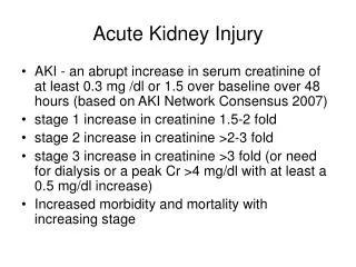
AKIN criteria • The Acute Kidney Injury Network (AKIN) modified the RIFLE criteria in order to: • include less severe ARF, • impose a time constraint of 48 hours, • allow for correction of volume status and obstructive causes of ARF prior to classification. • The AKIN proposed the term acute kidney injury (AKI) to represent the entire spectrum of acute renal failure, recognizing that an acute decline in kidney function is often secondary to an injury that causes functional or structural changes in the kidneys and that the injury can have important consequences for the patient even if it does not lead to organ failure and a requirement for renal replacement therapy
The AKIN defined AKI as “an abrupt (within 48 hours) reduction in kidney function, currently defined as an absolute increase in serum creatinine level of more than or equal to 0.3 mg/dL (26.4 μmol/L), a percentage increase in serum creatinine of more than or equal to 50% (1.5-fold from baseline), or a reduction in urine output (documented oliguria of less than 0.5 mL/kg/hr for more than 6 hours).”
AKIN criteria • SCr: Serum creatinine. Only one criterion must be met of either the SCr criteria or the urine output criteria; if both are present, the criterion which places the patient in the higher stage of AKI is selected. • The diagnostic criteria should only be applied after volume status has been optimized. Only one criterion (creatinine or urine output) has to be fulfilled to qualify for a stage. • Urinary tract obstruction needed to be excluded if oliguria was used as the sole diagnostic criterion. • Individuals who receive RRT are considered to have met the criteria for stage 3 irrespective of the stage they are in at the time of RRT.
Prognosis • In patients with AKI, the chances of renal recovery and survival depend on: • the underlying etiology, • the duration of AKI • associated comorbidities. • There is increasing recognition that AKI is associated with an increased risk of dying even after discharge from hospital. • AKI due to ATN is usually reversible. However, several reports have highlighted an association between AKI and subsequent risk of developing CKD, even if the episode of AKI resolves and serum creatinine returns to baseline.
Risk Factors The exact etiology of AKI is not always obvious and occasionally more than one factor contributes to its development.
Pathophysiology • There are typically three categories of AKI: • Prerenal AKI • Intrensic AKI • Postrenal AKI • The pathophysiologic mechanisms differ for each of the categories.
Compensatory hormonal mechanisms of decreased renal perfusion.
Intrarenal AKI = Intrinsic renal failure • Caused by diseases that can affect the integrity of the tubules, glomerulus, interstitium, or blood vessels. Damage is within the kidney; changes in kidney structure can be seen on microscopy. • The most common cause of intrinsic renal failure is Acute Tubular Necrosis (ATN) and it accounts for approximately 50% of all cases of AKI. • ATN represents a pathophysiologic condition that results from toxic (aminoglycosides, contrast agents, or amphotericin B) or ischemic insult to the kidney. • ATN results in necrosis of the proximal tubule epithelium and basement membrane, decreased glomerular capillary permeability, and backleak of glomerularfiltrate into the venous circulation. • Maintenance of ATN is mediated by intrarenal vasoconstriction. • Glomerular, interstitial, and blood vessel diseases may also lead to intrinsic AKI, but occur with a much lower incidence. Examples include glomerulonephritis, systemic lupus erythematosus, interstitial nephritis, and vasculitis. • In addition, prerenal AKI can progress to intrinsic AKI if the underlying condition is not promptly corrected.
Drug Induced AKI - Homework • Discuss the mechanism of nephrotoxicity of the following drugs: • Aminoglycosides • Amphotericin B • Radiocontrast media • Cyclosporine and Tacrolimus • Angiotensin-Converting Enzyme Inhibitors and Angiotensin Receptor Blockers • Non-steroidal Anti-Inflammatory Drugs
TABLE 55-1. Drug-Induced Renal Structural-Functional Alterationsand Examples
Signs and Symptoms of Uremia • Peripheral edema • Weight gain • Nausea/vomiting/diarrhea/anorexia • Mental status changes • Fatigue • Shortness of breath • Pruritus • Volume depletion (prerenal AKI) • Weight loss (prerenal AKI) • Anuria alternating with polyuria (postrenal AKI) • Colicky abdominal pain radiating from flank to groin (postrenal AKI)
Physical Examination Findings • Hypertension • Jugular venous distention • Pulmonary edema • Rales • Asterixis • Pericardial or pleural friction rub • Hypotension/orthostatic hypotension (prerenal AKI) • Rash (acute interstitial nephritis) • Bladder distention (postrenal bladder outlet obstruction) • Prostatic enlargement (postrenal AKI)
Results of urinalysis and sodium, urea, blood urea nitrogen, and creatinine measurements in acute kidney injury
Desired Outcomes and Goals • A primary goal of therapy is ameliorating any identifiable underlying causes of AKI such as hypovolemia, nephrotoxic drug administration, or ureter obstruction. • Prerenal and postrenal AKI can be reversed if the underlying problem is promptly identified and corrected, while treatment of intrinsic renal failure is more supportive in nature. • There is no evidence that drug therapy hastens patient recovery in AKI, decreases length of hospitalization, or improves survival.
The evaluation and initial management of patients with acute kidney injury (AKI) should include: • an assessment of the contributing causes of the kidney injury, • an assessment of the clinical course including comorbidities, • a careful assessment of volume status, • the institution of appropriate therapeutic measures designed to reverse or prevent worsening of functional or structural kidney abnormalities.
Things you will be asked about or will need to watch for in practice: • How can ARF be prevented? • Is the ARF drug-induced? • How should ARF be treated? • How should drugs be dosed in ARF?
Prevention of AKI • The best preventive measure for AKI, especially in individuals at high risk, is to avoid medications that are known to precipitate AKI. • Nephrotoxicity is a significant side effect of aminoglycosides, angiotensin-converting enzyme inhibitors, angiotensin receptor antagonists, amphotericin B, nonsteroidal anti-inflammatory drugs, cyclosporine, tacrolimus, and radiographic contrast agents. • Unfortunately, an effective, non-nephrotoxic alternative may not always be appropriate for a given patient and the risks and benefits of selecting a drug with nephrotoxic potential must be considered. • For example, serious gram-negative infections may require double antibiotic coverage, and based on culture and sensitivity reports, aminoglycoside therapy may be necessary. In cases such as this, other measures to reduce the risk of AKI should be instituted. • Thus, identifying patients at high risk for development of AKI and implementing preventive methods to decrease its occurrence or severity is critical.
FIGURE 55-3. Recommended Interventions for Prevention of Contrast Nephrotoxicity
Is the ARF drug-induced? • Drug list: if drug-induced, remember to remove offending agent until urine flow is re-established.
How should ARF be treated? • options are limited to: • 1. prevention of adverse drug reactions by discontinuing nephrotoxic drugs/treat cause • 2. adjustment of drug dosages based on the patient’s renal function is desired. • 3. supportive therapy, such as fluid, 4. electrolyte, and nutritional support, • 5. renal replacement therapy (RRT), • 6. treatment of non-renal complications such as sepsis and gastrointestinal bleeding while regeneration of the renal epithelium occurs.
How should ARF be treated? What to check: . Volume status • BP. hydration is key when BP low; avoid diuretics until BP normalized. Symptomatic orthostasis is a systolic drop of 30 or a diastolic drop of 10 and indicates moderate to severe volume depletion in patients not on a rate controller. Patients on a rate controller can be orthostatic with smaller volume loss. • HR: an increase in heart rate with little change in blood pressure can indicate mild dehydration. Symptomatic tachycardia will indicate at least moderate dehydration. HR inc 30 • Weight. In mild volume depletion, 2-3% body weight lost. In moderate-severe volume loss, ≥ 5 % body weight lost. BUN:Cr ratio. A BUN:serum creatinine ratio of ≥ 20:1 indicates at least moderate volume depletion .