Download
1 / 12
130 likes | 344 Vues
Basic Diagnostic Methods in Medicine. Ján Jakuš. Classification. A. Biochemical Methods- Blood tests, Enzyme tests, Mineral content tests, Test for evaluation of concentration -Na, K, Ca..., Glucose, Urea, in blood plasma, in liquor or in urine....

E N D
Basic Diagnostic Methods in Medicine Ján Jakuš
Classification A. Biochemical Methods-Blood tests, Enzyme tests, Mineral content tests, Test for evaluation of concentration -Na, K, Ca..., Glucose, Urea, in blood plasma, in liquor or in urine.... B. Physical Methods 1. Mechanical : e.g. Ausculta-tion, Percussion, Palpation, Blood pressure (non-direct)method, Body temperature measurement.. 2. Electric: ECG, EEG, EMG, ENG, ERG, Audiometry, Blood pressure (direct method,) Blood flow, and Air flow tests, Chronaxi-metry ... 3. Electromechanical:Spiro-metry, Energymetry,Test for sceletal muscle con-tractions, the lenght and tonus evaluation...
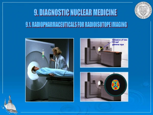
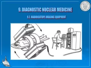
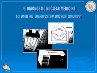
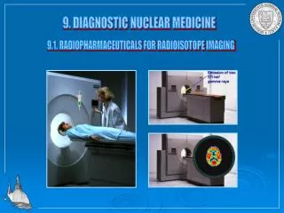
4. Optic and Optoelectric methods: Light microscopy, Electron microscopy,Ophtalmoscopy, Otoscopy, Bronchoscopy,Fiber optics... 5. Ultrasound (Dopplers)methods:Blood flow test, Sonography, Echocardiography... 6. X- ray Imagine methods: Sciascopy, Sciagraphy, Classic Tomography, Computer tomo-graphy (CT)... 7. Methods of Nuclear Medicine: Radioi-sotopes , Gammagraphy, Pozitron Emmision Tomography (PET).... 8. Magnetic scanning methods- Nuclear Magnetic Resonance Tomography (NMRT) 9.Combination of methods: AB and -8
Mechanical Methods Palpation- is a kind of an old, and subjective meth-od for evaluation of size and shape of organs within a body (e.g. liver, kidney, spleen, lymphatic glands, appendix bowel... Percussion- this old and subjective method- It looks for size and shape of organs and their boundaries (e.g. lungs, heart).Doctor uses his 3rd digit in order to strike the skin above the organ. As a result are different kinds of sounds Auscultation- old and subjective method looking for sounds and murmours determined by stethoscope Blood pressure and Temperature measurements(seepracticals)
Electrical Methods - ECG,EEG,EMG,ENG Electrocardiography (ECG)-method for record of heart electric signal from the surface of theskin. (For explanation of ECG curve and technique of re-cording see Practicals and Biophysical elixir) Electroencphalography (EEG)- a method for record -ing of brain electric signal from the scalp.The po-int is evaluation of frequency (f) and amplitude(A) of waves e.g. in Epilepsy. Waves or Rhythms: Alphaare recorded at rest with closed eylids with f = 8-13 Hz and A = 50 μV. Beta–when eyelids are open,f =15-20Hz, A = 5-10 μV Theta –pathology in adults,f = 4-7 Hz, A = 50 μV Delta- at REM Sleep f = 1- 4 Hz, A = 100 μV
Electrocardiography ECG leads Bipolar: I.II.III, CR, Cl, CF Unipolar: VR,VL,VF, aVR, aVL, aVF, V1-V6) ECG curve
Optic and Optoelectric Methods Light Microscopy- uses visible light. Microscop consists of ocular, objective,condenser, lateral and angular drift. Microscop increases the angle between two dots, thereby to percieve them as a two. Resolution :10-4-10-7 m (1/10 mm- 1/10 nm. Electron Microscopy- uses flow of electrons. Their source is „electron gun“., then pass through very thin layer of explored tissue and finally reach the projective . Picture is displayed on monitor. Reso-lution10-6 – 10-9m (from μm to ηm) Fiber optics- consists of130 cm long tube, with ca-nals: canal for image formation, canal for light, , working canal, rinsing canal.Doctor looks through an ocular in order to see the image of failured organ (e.g.stomach ulcer, tumors, etc).
Ultrasound - Dopplers methods The ultrasound(US)is a sound with f >20kHz (MHz) Sources: piesoelectric crystals, generators of US Point: US targets body, one part of it is absorbedby tissues and another part is refracted back to the probe of of the piesocrystal (Dopplers effect) Refracted US is named ECHOE Rule:The higher is US frequency (Mhz), the lower is it penetration, but better is a resolution of the org-an and vice versa. ECHOESare detected by spe-cial sensors and processed and displayed on black - white or colour monitor. US methods (So-nography, Echocardiography, Angiography) US is harmless,safe and very useful non-invasive methods. (use in pregnant women )