Download
1 / 37
370 likes | 387 Vues
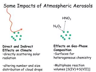
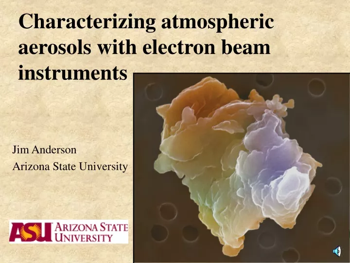
Characterizing atmospheric aerosols with electron beam instruments. Jim Anderson Arizona State University. Material presented here from research in collaboration with: Peter Crozier Cindy Twohy Hua Xin. Basic electron beam tools:. Scanning electron microscope (SEM).

E N D
Characterizing atmospheric aerosols with electron beam instruments Jim Anderson Arizona State University
Material presented here from research in collaboration with: Peter Crozier Cindy Twohy Hua Xin
Basic electron beam tools: Scanning electron microscope (SEM) [different SEMs for different purposes]
Transmission electron microscope (TEM) [different TEMs for different purposes]
Interaction of primary electron beam with atoms produces a number of useful signals Imaging of surface topography - secondary electrons (SEM) Imaging of internal structure - transmitted primary electrons (TEM) Imaging of average atomic number - backscattered electrons (SEM) Analysis of elemental composition - (1) characteristic X-rays by energy-dispersive spectroscopy (EDS) (SEM and TEM) (2) electron energy loss spectroscopy (EELS) - (TEM)
3 nm < 500 nm > TEM imaging of INDOEX ambient aerosols
Sea salt particle from RICO C-130 RF6 Intensity X-ray energy
rsj42202-1.2.1.eds Filter Fit Method Chi-sqd = 385.88 Livetime = 60.0 Sec. Beam Current = 400.000 nA ZAF Correction Acc.Volt.= 15 kV Take-off Angle=35.00 deg Number of Iterations = 4 Element k-ratio ZAF Atom % Element Wt % Err. Wt % (1-Sigma) Na-K 0.0025 1.624 6.02 0.41 +/- 0.05 Mg-K 0.0006 1.374 1.16 0.08 +/- 0.03 Al-K 0.0191 1.252 30.00 2.39 +/- 0.04 Si-K 0.0275 1.393 46.28 3.84 +/- 0.06 P -K 0.0002 1.694 0.36 0.03 +/- 0.04 S -K 0.0005 1.429 0.76 0.07 +/- 0.04 Cl-K 0.0004 1.348 0.47 0.05 +/- 0.04 K -K 0.0076 1.174 7.72 0.89 +/- 0.06 Ca-K 0.0005 1.133 0.49 0.06 +/- 0.05 Ti-K 0.0008 1.176 0.65 0.09 +/- 0.05 Cr-K 0.0000 1.125 0.00 0.00 +/- 0.00 Mn-K 0.0000 1.151 0.00 0.00 +/- 0.00 Fe-K 0.0072 1.124 4.9 0.81 +/- 0.16 Br-L 0.0000 1.244 0.00 0.00 +/- 0.00 Ba-L 0.0000 1.352 0.0 0.00 +/- 0.00 Pb-M 0.0000 1.635 0.00 0.00 +/- 0.00 Ni-K 0.0001 1.106 0.03 0.01 +/- 0.09 As-L 0.0000 1.346 0.00 0.00 +/- 0.00 Sn-L 0.0017 1.320 0.64 0.22 +/- 0.17 Sb-L 0.0005 1.315 0.19 0.07 +/- 0.11 Sr-L 0.0000 1.440 0.00 0.00 +/- 0.00 Zn-L 0.0000 1.539 0.00 0.00 +/- 0.00 Cu-K 0.0004 1.162 0.27 0.05 +/- 0.13 Total 100.00 9.08 Correction of X-ray data from irregular particles poses some problems. Aggregation makes the problems worse. However, when dealing with analyses from large populations subjected to cluster analysis, the problems tend to cancel out.
400 nm Position (nm) EELS Relative concentrations of carbon (open diamonds), nitrogen(solid squares) and sulphur (open triangles) as functions of position (in nanometers) from organic particle (INDOEX, cloud droplet residue from CVI).
Automated SEM analysis - characterization of size, shape , and composition of large numbers of particles (typically about 1000 per sample). SW Phoenix ambient aerosols
Undeliquesced particle shapes: What modelers requested... …what the atmosphere provided.
From PELTI - circularity distribution for an African dust sample. SW Phoenix dust
ACE-2 Sea salt aerosols
< 200nm > Ammonium sulfate
Some strictly preliminary results from December RICO samples -
Giant nucleii slides 56 microns width of field