Download
1 / 16
220 likes | 759 Vues
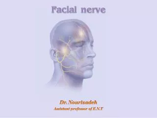
CN VII: FACIAL NERVE. Idham Hafize Supi 1090033 Nurmarzura Abdul Latif 1090045. I ntroduction. The facial nerve is the seventh cranial nerve, or simply cranial nerve VII.

E N D
CN VII: FACIAL NERVE IdhamHafizeSupi 1090033 Nurmarzura Abdul Latif1090045
Introduction • The facial nerve is the seventh cranial nerve, or simply cranial nerve VII. • It emerges from the brainstem between the pons and the medulla, and controls the muscles of facial expression, and functions in the conveyance of taste sensations from the anterior two-thirds of the tongue and oral cavity. • It also supplies preganglionic parasymphatetic fibers to several head and neck ganglia
Anatomy • The motor part of the facial nerve arises from the facial nerve nucleus in the pons while the sensory and parasympathetic parts of the facial nerve arise from the nervus intermedius. • The facial nerve eventually exits via the stylomastoid foramen to enter into the parotid where it branches into its peripheral segments. • The motor part and sensory part of the facial nerve enters the petrous temporal bone via the internal auditory meatus (intimately close to the inner ear) then runs a tortuous course (including two tight turns) through the facial canal, emerges from the stylomastoid foramen and passes through the parotid gland, where it divides into five major branches. Though it passes through the parotid gland, it does not innervate the gland. • The facial nerve forms the geniculate ganglion within the facial canal at the genu, the first bend in the canal.
Intracranial branches • Greater petrosal nerve - provides parasympathetic innervation to several glands, including the nasal gland, palatine gland, lacrimal gland, and pharyngeal gland. It also provides parasympathetic innervation to the sphenoid sinus, frontal sinus, maxillary sinus, ethmoid sinus and nasal cavity. • Nerve to stapedius - provides motor innervation for stapedius muscle in middle ear. • Chorda tympani • Submandibular gland • Sublingual gland • Special sensory taste fibers for the anterior 2/3 of the tongue.
Extracranial branches Distal to stylomastoid foramen, the following nerves branch off the facial n: • Posterior auricular nerve - controls movements of some of the scalp muscles around the ear • Branch to Posterior belly of Digastric muscle as well as the Stylohyoid muscle • Five major facial branches (in parotid gland) - from top to bottom (Tolong Zip Baju Makcik Cepat): • Temporal branch of the facial nerve • Zygomatic branch of the facial nerve • Buccal branch of the facial nerve • Marginal mandibular branch of the facial nerve • Cervical branch of the facial nerve
Function • Other • The facial nerve also supplies parasympathetic fibers to the submandibular gland and sublingual glands via chorda tympani. Parasympathetic innervation serves to increase the flow of saliva from these glands. It also supplies parasympathetic innervation to the nasal mucosa and the lacrimal gland via the pterygopalatine ganglion. • The facial nerve also functions as the efferent limb of the corneal reflex.
Clinical examination • Motor:-muscle of facial expression • This nerve is therefore tested by asking the patient to crease up their forehead (raise their eyebrows), close their eyes and keep them closed against resistance, puff out their cheeks and reveal their teeth. • Sensory:-taste • The four primary tastes are bitter, sweet, sour, and salty. Screen for disorders of sweet or salty taste with salt and sugar. • With the patient's eyes closed and tongue protruded, take a tongue blade and smear a small amount of salt or sugar on the lateral surface and side of the tongue. • Instruct the patient to tell you the identity of the substance. Rinse the mouth thoroughly and repeat the test on the other side, using a different substance.
Abnormalities:- Intracranial lesions • Intracranial lesions occur during the intracranial course of the facial nerve (proximal to the stylomastoid foramen). • The muscles of facial expression will be paralysed or severely weakened. • The other symptoms produced depend on the location of the lesion, and the branches that are affected: • Chordatympani – reduced salivation and loss of taste on the ipsilateral 2/3 of the tongue. • Nerve to stapedius – ipsilateralhyperacusis (hypersensitive to sound). • Greater petrosal nerve – ipsilateral reduced lacrimal fluid production. • The most common cause of an intracranial lesion of the facial nerve is middle ear pathology – such as a tumour or infection. If no definitive cause can be found, the disease is termed Bell’s palsy.
Extracranial lesions • Extracranial lesions occur during the extracranial course of the facial nerve (distal to the stylomastoid foramen). • Only the motor function of the facial nerve is affected, resulting in paralysis or severe weakness of the muscles of facial expression. • There are various causes of extracranial lesions of the facial nerve: • Parotid gland pathology – e.g a tumour. • Infection of the nerve- particularly by the herpes virus. • Compression during forceps delivery – the neonatal mastoid process is not fully developed, and does not provide complete protection of the nerve. • Ideopathic – If no definitive cause can be found, the disease is termed Bell’s palsy.