Download
1 / 39
440 likes | 928 Vues
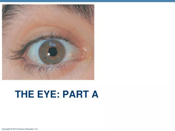
The eye: part a. The Eye and Vision. 70% of all sensory receptors are in the eye Nearly half of the cerebral cortex is involved in processing visual information! Most of the eye is protected by a cushion of fat and the bony orbit. Eyebrow. Eyelid. Eyelashes. Site where conjunctiva

E N D
The Eye and Vision • 70% of all sensory receptors are in the eye • Nearly half of the cerebral cortex is involved in processing visual information! • Most of the eye is protected by a cushion of fat and the bony orbit
Eyebrow Eyelid Eyelashes Site where conjunctiva merges with cornea Palpebral fissure Lateral commissure Iris Eyelid Sclera (covered by conjunctiva) Lacrimal caruncle Pupil Medial commissure (a) Surface anatomy of the right eye Figure 15.1a
Eyebrows • Eyebrows-Overlie the supraorbital margins • Shade the eye • Prevent perspiration from reaching eye
Eyelids (Palpebrae) • Protect the eye anteriorly • Levator palpebrae superioris muscle—gives upper eyelid mobility • Tarsal plates—internal supporting CT sheet • Palpebral fissure—separates eyelids • Medial and Lateral Canthi – eye angles • Caruncle—fleshy elevation at medial canthus • Contains oil and sweat glands • Eyelashes • Nerve endings of follicles initiate reflex blinking
Site where conjunctiva merges with cornea Palpebral fissure Lateral commissure Eyelid Sclera (covered by conjunctiva) Lacrimal caruncle Pupil Medial commissure (a) Surface anatomy of the right eye Figure 15.1a
Eyelids • Tarsal (Meibomian) Glands • Modified sebaceous glands • Produce oily secretion; prevents eyelids from sticking, lubricates eyelid • Embedded in tarsal plates • Ducts open posterior to eyelashes
Eyelids • Ciliary Glands • Sebaceous and sweat glands • Found between eyelashes
Conjunctiva • Transparent mucous membrane • Palpebral conjunctiva: lines eyelids • Bulbar conjunctiva: covers white of the eyes anteriorly
Levator palpebrae superioris muscle Orbicularis oculi muscle Tarsal plate Palpebral conjunctiva Tarsal glands Cornea Palpebral fissure Bulbar conjunctiva Orbicularis oculi muscle (b) Lateral view; some structures shown in sagittal section
Lacrimal Apparatus • Consists of: Lacrimal gland and ducts that connect to nasal cavity • Releases: Lacrimal secretion (tears) • The solution also contains: mucus, antibodies, and lysozyme (bacteria destroying enzyme) • The solution drains via: paired lacrimal puncta into lacrimal canaliculi then into nasolacrimal duct
Lacrimal sac Lacrimal gland Excretory ducts of lacrimal glands Lacrimal punctum Lacrimal canaliculus Nasolacrimal duct Inferior meatus of nasal cavity Nostril Figure 15.2
Extrinsic Eye Muscles • The movement of the eye is controlled by six muscles on the external surface of each eye • Superior rectus: elevates eye • Inferior rectus: depresses eye • Lateral rectus: moves eye laterally • Medial rectus: moves eye medially • Inferior oblique: Moves eye up and out • Superior oblique: Moves eye down and out
Superior oblique muscle Superior oblique tendon Superior rectus muscle Lateral rectus muscle Inferior rectus muscle Inferior oblique muscle (a) Lateral view of the right eye Figure 15.3a
Trochlea Superior oblique muscle Superior oblique tendon Axis at center of eye Superior rectus muscle Inferior rectus muscle Medial rectus muscle Lateral rectus muscle Common tendinous ring (b) Superior view of the right eye Figure 15.3b
Extrinsic Eye Muscles • The innervation to each muscle can be remembered by the following equation: (LR6SO4)O3 • Which means: • Lateral rectus: is innervated by CN # 6 (Abducens) • Superior oblique: is innervated by CN #4 (Trochlear) • All others: are innervated by CN #3 (oculomotor)
Structure of the Eyeball • Wall of eyeball contains three layers (tunics) • Fibrous layer - Outermost layer; dense avascular CT with two regions: sclera and cornea • Vascular layer - Middle pigmented layer with three regions: choroid, ciliary body, and iris • Sensory layer (retina) -Delicate two-layered membrane
Anterior segment (contains aqueous humor) Lens Posterior segment (contains vitreous humor)
Structures or Substances to Know: • Sclera • Cornea • Choroid • Ciliary body, muscle and processes • Suspensory ligaments • Ora Serrata • Iris • Pupil • Anterior segment- divided into anterior and posterior chambers • Posterior segment • Vitreous humor • Aqueous humor • Lens • Macula Lutea • Fovea Centralis • Optic disc • Optic nerve • Central artery and vein • Canal of Schlemm
Parasympathetic + Sympathetic + Iris (two muscles) • Sphincter pupillae • Dilator pupillae Dilator muscle contraction increases pupil size. Pupillary Sphincter muscle contraction decreases pupil size. Figure 15.5
Ciliary body Ciliary zonule (suspensory ligament) Sclera Choroid Iris Pupil Optic nerve Lens Optic disc (blind spot) Figure 15.4a
Sensory Layer: Retina • Delicate two-layered membrane • Pigmented layer • Outer layer • Absorbs light and prevents its scattering • Stores vitamin A
Sensory Layer: Retina • Neural layer • Photoreceptor cells: transduce light energy • Cells that transmit and process signals: bipolar cells, ganglion cells, amacrine cells, and horizontal cells
Pathway of light Neural layer of retina Pigmented layer of retina Choroid Sclera Optic disc Central artery and vein of retina Optic nerve (a) Posterior aspect of the eyeball Figure 15.6a
The Retina • Ganglion cell axons • Run along the inner surface of the retina • Axons of ganglion cells leave eye as the optic nerve • Optic disc (blind spot) • Site where the optic nerve leaves the eye • Lacks photoreceptors
Pathway of light Neural layer of retina Pigmented layer of retina Choroid Sclera Optic disc Central artery and vein of retina Optic nerve (a) Posterior aspect of the eyeball Figure 15.6a
Photoreceptors • Rods • More numerous at peripheral region of retina, away from the macula lutea • Operate in dim light • Provide indistinct, fuzzy, non color peripheral vision
Photoreceptors • Cones • Found in the macula lutea; concentrated in the fovea centralis • Operate in bright light • Provide high-acuity color vision
Photoreceptors Bipolar cells • Rod Ganglion cells • Cone Amacrine cell Horizontal cell Pathway of signal output Pigmented layer of retina Pathway of light (b) Cells of the neural layer of the retina Figure 15.6b
Blood Supply to the Retina • Two sources of blood supply • Choroid supplies: the outer third (photoreceptors) • Central artery and vein of the retina supply: the inner two-thirds
Central artery and vein emerging from the optic disc Macula lutea Optic disc Retina Figure 15.7
Internal Chambers and Fluids • The lens and ciliary zonule separate the anterior and posterior segments
Ciliary body Ciliary zonule (suspensory ligament) Sclera Choroid Retina Macula lutea Fovea centralis Posterior pole Pupil Optic nerve Anterior segment (contains aqueous humor) Lens Posterior segment (contains vitreous humor) Optic disc (blind spot) Figure 15.4a
Internal Chambers and Fluids • Posterior segment contains vitreous humor that: • Transmits light • Supports posterior surface of the lens • Holds the neural retina firmly against the pigmented layer • Contributes to intraocular pressure • Anterior segment is composed of two chambers • Anterior chamber—between the cornea and the iris • Posterior chamber—between the iris and the lens
Internal Chambers and Fluids • Anterior segment contains aqueous humor • Plasma like fluid continuously filtered from capillaries of the ciliary processes • Drains via the scleral venous sinus (canal of Schlemm) at the sclera-cornea junction • Supplies: nutrients and oxygen mainly to the lens and cornea but also to the retina, and removes wastes • Glaucoma: compression of the retina and optic nerve if drainage of aqueous humor is blocked
Iris Posterior segment (contains vitreous humor) Lens Cornea 2 Ciliary zonule (suspensory ligament) Aqueous humor Anterior chamber Anterior segment (contains aqueous humor) Posterior chamber Ciliary body 3 1 Scleral venous sinus Ciliary processes Corneal- scleral junction Ciliary muscle Cornea Lens Figure 15.8
Lens • Biconvex, transparent, flexible, elastic, and avascular • Allows precise focusing of light on the retina • Cells of lens epithelium differentiate into lens fibers that form the bulk of the lens • Lens fibers—cells filled with the transparent protein crystallin • Lens becomes denser, more convex, and less elastic with age • Cataracts (clouding of lens) occur as a consequence of aging, diabetes mellitus, heavy smoking, and frequent exposure to intense sunlight
Ora serrata Ciliary body Ciliary zonule (suspensory ligament) Sclera Choroid Retina Cornea Macula lutea Iris Fovea centralis Posterior pole Pupil Optic nerve Anterior pole Anterior segment (contains aqueous humor) Lens Scleral venous sinus Central artery and vein of the retina Posterior segment (contains vitreous humor) Optic disc (blind spot) (a) Diagrammatic view. The vitreoushumor is illustrated only in thebottom part of the eyeball. Figure 15.4a