Download
1 / 48
800 likes | 1.63k Vues
Corticosteroid - Induced Osteoporosis. Chatlert Pongchaiyakul. MD. Endocrinology Unit, Medicine Department Faculty of Medicine, Khon Kaen University. . Osteoporosis. Systemic skeletal disease Low bone mass Microarchitectural deterioration of bone tissue

E N D
Corticosteroid - Induced Osteoporosis Chatlert Pongchaiyakul. MD. Endocrinology Unit, Medicine Department Faculty of Medicine, Khon Kaen University.
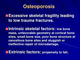
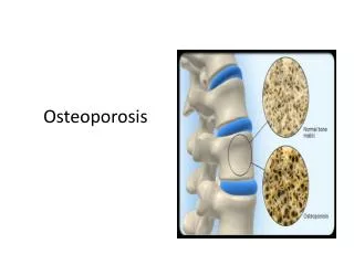
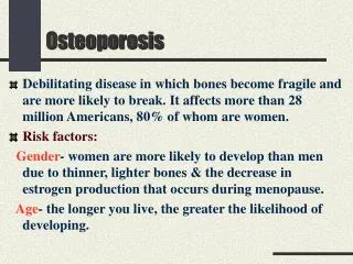
Osteoporosis • Systemic skeletal disease • Low bone mass • Microarchitectural deterioration of bone tissue • Increase in bone fragility and fracture susceptibility
Clinical Burden of CIO • Most common form of drug-related osteoporosis in men and women • Occurs at any age, in both genders, across races • Up to 50% of patients on chronic steroid therapy sustain osteoporotic fractures and/or develop osteonecrosis
Corticosteroid-Induced Osteoporosis • Common, iatrogenic form of secondary osteoporosis • Associated with corticosteroid use in chronic, noninfectious medical conditions • Asthma - Nephrotic syndrome • Chronic lung disease - Transplantation • Rheumatologic disorders - etc • Inflammatory bowel disease
Clinical significant - Increase bone loss and fracture : 6 Mo. - Trabecular > cortical bone - 7.5 mg of prednisolone ( equivalent ) - Incidence of osteoporosis ~ 30-50% - Vertebral fracture 30-35 % , hip fracture 50% - Rate of bone loss 2-4 % per year - Alternate day regimen , inhale steroids
Fracture Risk and Dose of Corticosteroids 6 5 4 Relative risk of fracture compared with control Hip fracture 3 Vertebral fracture 2 1 0 2.5 mg/d 2.5-7.5 mg/d >7.5 mg/d Relative risk of fracture by dosages of corticosteroids of prednisolone. van Staa TP, et al, 1998.
120 100 r=-0.39 (P=0.009) 80 Percent predicted bone density 60 40 2 4 6 8 10 12 14 16 18 20 22 24 26 28 30 32 34 36 Duration of corticosteroid use (years) CIO in Patients With Asthma Relationship of percentage predicted bone density to duration of corticosteroid use in 44 corticosteroid-treated asthmatic patients. Schatz M, Dudl J, Zeiger RS, et al. Allergy Proc. 1993;14:341-345. Reprinted with permission.
CIO in Patients With Rheumatoid Arthritis CS=corticosteroid; therapy = 7 mg prednisone equivalent per day. Density change measured as change in absolute or Z score (difference in standard deviation compared with healthy age-matched controls of the same race and sex) compared to baseline. Verhoeven AC, et al, 1997.
CIO and Systemic Lupus Erythematosus * ** * ** *P<0.001; **P=0.002. Percentage of SLE patients (N=97) with low BMD, as measured by DXA. Kipen Y, et al, 1997.
Potential Factors Causing Bone Loss in Inflammatory Bowel Disease • Corticosteroids • Vitamin D / Calcium deficiency • Poor nutritional status • Inflammation • Physical inactivity • Concurrent medications (immunosuppressive agents)
CIO and Chronic Obstructive Pulmonary Disease * ** *P<0.05 vs. ISU or NSU; **P<0.005 vs ISU. McEvoy CE, et al, 1998.
Pathophysiology of CIO: Overview • Bone remodeling occurs throughout adulthood • Osteoporosis results from an imbalance between osteoclast and osteoblast activity • Two metabolic abnormalities contribute to increased bone resorption • Secondary hyperparathyroidism due to decreased GI absorption and urinary excretion of calcium • Altered gonadal function and decreased adrenal production of androgens
Pathophysiology of CIO Calcium homeostasis Gonadal hormone Inhibit bone formation Increase bone resorption other
Calcium homeostasis Decrease calcium and phosphate from GI tracts unknown mechanism Increase urinary calcium excretion decrease calcium reabsorption at distal tubules Stimulatiom PTH secretion
Gonadal hormone effects Decrease sex hormone : direct & indirect Decrease LH from pituitary gland : estrogen and testosterone Decrease synthesis from adrenal glands Decrease sex hormone binding globulin
Bone formation and bone resorption Osteoblast - inh. Osteoblast proliferation - decrease matrix synthesis - increase apoptosis - decrease protein synthesis ( type 1 collagen and noncollagenous protein - decrease osteocalcin , IGF1, IGFBP3,5 , insulin-like growth factors, transforming growth factor B , prostaglandin E
Bone formation and bone resorption Osteoclast increase osteoclast activity increase apoptosis of mature osteoclast
Osteoblast proliferation Apoptosis OB number Protein synthesis Bone formation Differentiation Bone mass Fracture Risk Androgen Osteoclast apoptosis Bone resorption Osteoclast formation PTH Calcium and phosphate absorption ( gut and kidney ) Glucocorticoid
Diagnosis of CIO: Initial Clinical Work-Up • Medical history • Risk factors for bone loss • Physical exam • Clinical signs and symptoms
Patient Evaluation History Documentation of height , weight , muscle strength , balance , vision Documentation of medical history Documentation of menstrual history, infertility in men Fracture history and Family history of fractures Other risk factors for osteoporosis : - Lifestyles influences : calcium and vitamin D intake, smoking, alcohol intake, medications, prevention of falling - Patient education : prevention of falling , exercise General health and prognosis
Patient Evaluation Physical examination Evidence of osteoporosis : evidence of fracture , kyphosis , loss of height , muscle strength and size General physical findings : assessment of underlying disorder , other medical conditions
Patient Evaluation laboratory Complete blood count and erythrocyte sedimentation rate ( ESR ) Serum calcium, phosphate, creatinine, electrolyte, alkaline phosphatase, 25-hydroxyvitamin D, estradiol, testosterone ( male ) 24 hr-Urinary calcium and creatinine BMD of spine and hip X-rays of appropriate areas
WHO Criteria for Assessing Disease Severity Diagnostic Criteria* Classification T= 0 to -1 SD Normal T= -1 to -2.5 SD Osteopenia T-2.5 SD Osteoporosis T-2.5 SD + fragility fractures Severe osteoporosis * Measured in “T scores,” ie, the number of standard deviations below or above the peak bone mass in a young adult reference population of the same sex; SD=standard deviation.
Guidelines for BMD Measurement • Baseline BMD prior to/within 6 months of initiating therapy • Antero-posterior measurement of lumbar spine and femoral neck • Follow-up at 6 and 12 months, annually thereafter until bone mass stabilizes • Measuring hip alone may miss more rapid loss in spine
Management of CIO: Goals of Treatment • Reduce fracture risk • Maintain current BMD, prevent additional bone loss • Alleviate pain associated with existing fracture(s) • Maintain/increase muscle strength • Initiate lifestyle changes as needed
BMD, Vitamin D, and Calcium 6 12 18 24 30 36 months months months months months months 0 -2 -4 from baseline (%) Change in lumbar spine BMD -6 -8 -10 -12 Vitamin D & calcium Placebo Adachi JD, et al, 1996.
Action • Inhibit bone resorption • Prevent apoptosis of osteoblasts • Partially reverse bone loss • Prevent early resorptive phase • of bone loss • Inhibit bone resorption • Maintain or increase bone mass Treatment Hormonal replacement therapy Calcitonin Bisphosphonates Pharmacologic Treatment of CIO: Overview
Pharmacologic treatment of CIO Thiazide diuretics increase calcium absorption from GI tract decrease urinary calcium excretion Fluorides stimulate osteoblast activity Anabolic steroids increase bone formation
Hormone Replacement Therapy in the Treatment of CIO: ACR Guidelines Patient group Postmenopausal women Premenopausal women w/intact ovarian functions (ages 13-50) Men Recommendation • Estrogen + progestin for women with intact uteri • Bisphosphonate or calcitonin if HRT contraindicated • Estrogen-containing OCs (50 g estradiol) or equivalent • Bisphosphonate or calcitonin ifestrogen contraindicated • Testosterone (if serum testosterone levels low) • Bisphosphonate or calcitonin if testosterone contraindicated American College of RheumatologyTask Force on Osteoporosis Guidelines, 1996.
Estrogen Replacement Therapy in the Treatment of CIO Group 1 Group 2 ) 2 Prednisone Prednisone Group 3 Group 4 only + ERT Control ERT only 0.06 * 0.04 Changes in lumbar spine BMD (g/cm 0.02 at 1 year 0 -0.02 -0.04 -0.06 *P=0.008 vs. baseline; P=0.027 between groups 1 and 2. Lukert BP, et al, 1992.
Testosterone Replacement Therapy in the Treatment of CIO * 5.0 2.5 Changes in lumbar spine BMD (%) at 1 year 0.0 Testosterone therapy Control period period -2.5 -5.0 *P=0.005 vs control; P=0.05 between-group difference. Reid IR, et al, 1996.
Cyclical Etidronate and Prevention of Corticosteroid-Induced Bone Loss Etidronate Control * 2 * 1 0 -1 -2 -3 Changes in BMD from baseline (%) at 1 year -4 Lumbar Femoral Trochanter Lumbar Femoral Trochanter spine neck spine neck *P<0.05 between-group difference. Adachi JD, et al, 1997. Roux C, et al,1998.
Etidronate: Pooled Results from Three Randomized Trials 6 4 Change in BMD from baseline (%) 2 0 Lumbar spine* Femoral neck Trochanter Men Pre-menopausal women Post-menopausal women *P<0.05 between-group difference. Roux C, et al,1998.
Efficacy of Pamidronate in the Prevention of Bone Loss 6 4 2 Changes in BMD from baseline (%) 0 -2 -4 -6 6 months 12 months 6 months 12 months Pamidronate + calcium Calcium only Boutsen Y, et al, 1997.
Control Alendronate 5 mg Alendronate 10 mg 3.5 2.5 Change in BMD from baseline (%) 1.5 at 48 weeks 0.5 -0.5 -1.5 Lumbar spine Femoral neck Trochanter Total body Efficacy of Alendronate in Increasing BMD * * * * ** * ** *P <0.001 vs. control; **P <0.01 vs. control; †P <0.001 vs. baseline, ‡P <0.01 vs. baseline; Saag KG, et al, 1998.
Efficacy of Alendronate: Two Years Follow-Up Control Alendronate 10 mg Alendronate 5 mg Alendronate 2.5 mg year 1, 10 mg year 2 † * 4 * † 3 2 1 Change in BMD from baseline (%) ** ** 0 -1 -2 -3 -4 Lumbar spine Femoral neck Trochanter *P<0.001 vs. control; **P<0.01 vs. control; †P<0.05 vs. control. Saag KG, et al, 1998.
Effect of Risedronate on BMD inPatients Initiating Corticosteroid Therapy Control Risedronate 2.5 mg 4.0 Risedronate 5 mg 2.0 * * * * at 12 months * * 0.0 Change in BMD from baseline (%) -2.0 -4.0 Lumbar spine Femoral neck Trochanter *P<0.05 vs control. Cohen S, et al, 1998.
Effect of Risedronate on BMD in Patients on Long-Term Corticosteroid Therapy * 3.0 * * 2.0 1.0 * at 12 months Change in BMD from baseline (%) 0.0 Lumbar spine Femoral neck Trochanter -1.0 -2.0 Control Risedronate 2.5 mg Risedronate 5 mg -3.0 *P<0.05 vs. control. Devogelaer JP, et al, 1998.
Effect of Risedronate on Vertebral Fracture Rates 20 15 10 Patients with vertebral fractures (%) * 5 0 Pooled control patients Pooled risedronate patients Pooled vertebral fracture rates from 518 patients on steroid therapy. *P=0.016 vs. control. Reid D, et al, 1998.
Bisphosphonates in the Management of CIO: A Meta-Analysis Treatment Number of Change in lumbar pooled trials spine BMD (%)* Vitamin D 18 +1.96 Calcitonin 11 +2.11 Bisphosphonates 18 +5.31† *Compared with no treatment or with calcium alone †P=0.0001 compared with calcitonin or vitamin D
Glucocorticoid therapy evaluation Plan- at start of glucocorticoid therapy 1. Minimize glucocorticoid dose 2. Use alternate day therapy , topical steroid or bone sparing steroid if possible 3. Prescribe exercise ( weight baring ) , physical therapy , prevent falling 4. Avoid smoking and excess alcohol 5. Assure adequate calcium intake 6. Add supplement calcium up to 1000-15000 mg calcium /day 7. Add multivitamin containing 400-800 IU vitamin D 8. BMD measurement of the spine and hip : if T-score lower than –1 SD start HRT and if more than –1 SD start HRT only in postmenopausal woman
Glucocorticoid therapy evaluation Reassessment at 2-3 mo 1. Review glucocorticoid therapy : attempt to decrease or discontinue 2. Assess exercise and calcium intake 3. Measure serum calcium , 24 hr urinary calcium if more than 4 mg/kg/d use hydrochlorothiazide 25-50 mg twice dailyReassessment at 6 mo 1. Review glucocorticoid therapy and minimize 2. Assess exercise and calcium intake 3. Repeat serum calcium and 24 hr urinary calcium measurement 4. Alter calcium / vitamin D / thiazide therapy if necessary 5. If pateint is to continue glucocorticoid ,consider to repeat BMD 6. Consider HRT / bisphosphonate/ calcitonin
Glucocorticoid therapy evaluation Reassessment at 1 yr 1. Review glucocorticoid therapy and minimize 2. Assess exercise and calcium intake 3. Repeat serum calcium and 24 hr urinary calcium measurement 4. BMD measurement ( spine and hip ) 5. Alter calcium / vitamin D / thiazide therapy if necessary 6. Alter further thereapy if bone loss if continues Reassessment thereafter if glucocorticoids continue 1. Repeat annual assessment as above 2. Change therapy as needed 3. Consider newer drugs as they become available
ACR Task Force on Osteoporosis: Initiating Long-Term Corticosteroid Therapy Initial history & physical, lab/DXA measurements Calcium/vitamin D supplementation Patient education T score < -1 Initiate HRT; bisphosphonates or calcitonin if HRT contraindicated T score > -1 Monitor regularly One month follow-up: Obtain 24h urine to measure calcium If > 300 mg/d: add thiazide diuretic Adjust dosage of calcium and vitamin D supplementation 6-12 months follow-up: Repeat BMD Decrease >5%: change/add medication Increase, no change, or decrease <5%: no change in therapy American College of Rheumatology Task Force on Osteoporosis Guidelines, 1996.
Anticipated therapy with glucocorticoid • Atraumatic fractures • Yes No • Calcium 1500 mg/day yes Measurement of bone mineral density • Vitamin D 400-800 IU/day Lower than 2SD below the mean for • Exercise >5 % young adults or Lower than 1 SD below the • Screen for hypogonadism bone loss mean for aged-match controls • No • If hypogonadism present : Calcium 1000 mg/day • Add hormone replacement with Vitamin D 400-800 IU/day • Estrogen in woman and testosterone in men Exercise • Check BMD in one year : add anti-resorptive Repeat bone mineral density in 1 yr. • Therapy if > 2 percent bone loss • If hypogonadism absent: < 5 % bone loss • Add bisphosphanate if no fracture pain • Add calcitonin if fracture pain • Continue conservative therapy as long as • bone density criteria above not met
Corticosteroid-Induced Osteoporosis: Conclusions • Most common form of drug-related osteoporosis • Imbalance in bone formation and resorption • Resultant bone loss and fracture • Bone densitometry is recommended for all patients on chronic steroid therapy • T scores -2.5 indicate osteoporosis • T scores -1 indicate osteopenia • Each standard deviation change in bone density is associated with at least a two-fold change in fracture risk
Corticosteroid-Induced Osteoporosis: Conclusions • Primary treatment goals • Reduce fracture risk • Maintain or increase bone mass • Vitamin D and calcium may slow early resorptive changes • HRT is recommended for patients with T scores <1 to prevent bone resorption (use bisphosphonates or calcitonin if HRT is contraindicated) • Bisphosphonates are an efficacious treatment • Inhibit bone resorption • Maintain or increase bone mass • Advanced generation bisphosphonates • Increase BMD of hip, spine, and total body • May lower risk for vertebral, hip, and forearm fractures
I will always... Love you with all my fractured bones.