Download
1 / 24
240 likes | 391 Vues
First week of Development. 2- Implantation. Dr Rania Gabr. Objectives. By the end of this lecture , the student should be able to: Define the term “implantation’. Describe the cleavage and the stage at which implantation occurs. Explain the process of blastocyst formation.

E N D
First week of Development 2- Implantation Dr Rania Gabr
Objectives • By the end of this lecture , the student should be able to: • Define the term “implantation’. • Describe the cleavage and the stage at which implantation occurs. • Explain the process of blastocyst formation. • Describe the process of ‘implantation’. • Define the normal site of implantation. • Describe the abnormal sites of implantation (ectopic pregnancy).
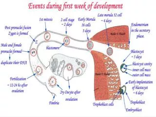
Cleavage Definition: It is the repeated mitotic divisions of the zygote, resulting into a rapid increase in the number of cells that are called blastomeres. Site: The uterine tube medial to the ampula.
Repeated mitoticdivisionsof the zygote. • Begins about 30 hoursafter fertilization. • There is rapid increase in the number of cells. • The cells (blastomeres) become smaller with each division. • Normally occurs as the zygote passes along the uterine tube to the uterus. • During cleavage, zygote is within the zona pellucida.
Stages of cleavage • After 8-cell stage, the cells become compactly arranged compaction • 12-16 cell stage is called MORULA. • It is formed about 3 days after fertilization and enters the uterine cavity.
Blastocyst formation: As the morulareaches the uterine cavity, fluid from the uterine cavity penetrates the zonapellucidaand accumulates in the inter-cellular spaces of the inner cell mass. -A single cavity is formed and called blastocele.
-The morula after the formation of blastocele is called blastocyst. • -The blastocyst consists of inner cell mass (it will form the future embryo; so it is called embryoblasts) and • outer cell mass (future trophoblast & placenta). • The zonapellucidadisappears allowing for implantation.
Blastocyst takes its nourishment from uterine secretions. • It enlarges in size. • It is ready to get attached and implanted to the uterine wall.
At this stage, the conceptus is called Blastocyst. It has two poles: embryonic & abembryonic.. Embryonic pole Abembryonic pole
Clinical application: • Embryonic stem cells (ES cells) are derived from the inner cell mass. • These cells can give any type of tissue. • So they are called pluripotent cells.
Implantation Definition: Penetration of the blastocyst into the superficial (compact) layer of the endometrium. The endometrium after implantation is called decidua. Time: Implantation occurs at the 6th dayafter fertilization and is completed about the 11th day.
6 days after fertilization: • Blastocyst attaches to the endometrial epithelium, usually adjacent to the embryonic pole.
Trophoblastproliferates rapidly and differentiates into two layers: 1-Inner cellular cytotrophoblast, and 2-Outer mass of syncytiotrophoblast(multinucleated protoplasm with no cell boundaries). Finger like processes of syncytiotrophoblastextend through the endometrium and invade the endometrial connective tissue.
By the end of 7th day, the blastocyst gets implanted in the superficial compact layer of the endometrium and derives its nourishment from the eroded endometrium.
The blastocyst gradually embed deeper in the endometrium and the defect in the endometrial epithelium is filled by closing plug (day 10).
The defect gradually disappear as the endometrial epithelium is repaired (day 12 & 13) Blood filled lacunae appear in syncytiotrophoblastwhich filled with maternal blood, establishing primitive uteroplacental circulation.
Site of Implantation: • The normal site is the endometrium of the posterior wall of the fundus of the uterus in or near the middle line. • Implantation in the lower segment leads to placenta praevia
Extrauterine: leading to ectopic pregnancies:1-fallopian tube2- ovary3- abdomen4- cervical