Download
1 / 88
900 likes | 1.25k Vues
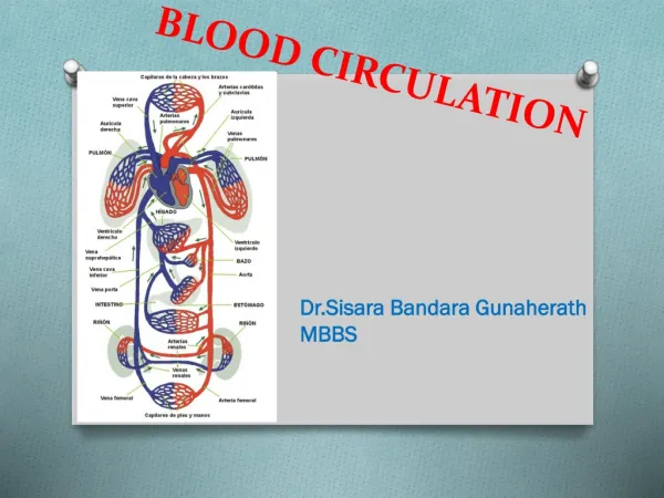
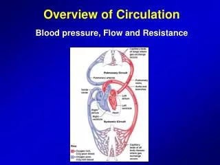
Overview of Blood Circulation. Blood leaves the heart via arteries that branch repeatedly until they become capillaries Oxygen (O 2 ) and nutrients diffuse across capillary walls and enter tissues Carbon dioxide (CO 2 ) and wastes move from tissues into the blood. Overview of Blood Circulation.

E N D
Overview of Blood Circulation • Blood leaves the heart via arteries that branch repeatedly until they become capillaries • Oxygen (O2) and nutrients diffuse across capillary walls and enter tissues • Carbon dioxide (CO2) and wastes move from tissues into the blood
Overview of Blood Circulation • Oxygen-deficient blood leaves the capillaries and flows in veins to the heart • This blood flows to the lungs where it releases CO2 and picks up O2 • The oxygen-rich blood returns to the heart
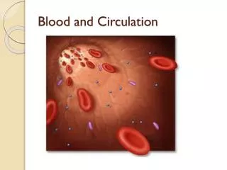
Composition of Blood • Blood is the body’s only fluid tissue • It is composed of liquid plasma and formed elements • Formed elements include: • Erythrocytes, or red blood cells (RBCs) • Leukocytes, or white blood cells (WBCs) • Platelets • Hematocrit – the percentage of RBCs out of the total blood volume
Components of Whole Blood Figure 17.1
Physical Characteristics and Volume • Blood is a sticky, opaque fluid with a metallic taste • Color varies from scarlet to dark red • The pH of blood is 7.35–7.45 • Temperature is 38C • Blood accounts for approximately 8% of body weight • Average volume: 5–6 L for males, and 4–5 L for females
Functions of Blood • Blood performs a number of functions dealing with: • Substance distribution • Regulation of blood levels of particular substances • Body protection
Distribution • Blood transports: • Oxygen from the lungs and nutrients from the digestive tract • Metabolic wastes from cells to the lungs and kidneys for elimination • Hormones from endocrine glands to target organs
Regulation • Blood maintains: • Appropriate body temperature by absorbing and distributing heat • Normal pH in body tissues using buffer systems • Adequate fluid volume in the circulatory system
Protection • Blood prevents blood loss by: • Activating plasma proteins and platelets • Initiating clot formation when a vessel is broken • Blood prevents infection by: • Synthesizing and utilizing antibodies • Activating complement proteins • Activating WBCs to defend the body against foreign invaders
Blood Plasma • Blood plasma contains over 100 solutes, including: • Proteins – albumin, globulins, clotting proteins, and others • Lactic acid, urea, creatinine • Organic nutrients – glucose, carbohydrates, amino acids • Electrolytes – sodium, potassium, calcium, chloride, bicarbonate • Respiratory gases – oxygen and carbon dioxide
Formed Elements • Erythrocytes, leukocytes, and platelets make up the formed elements • Only WBCs are complete cells • RBCs have no nuclei or organelles, and platelets are just cell fragments • Most formed elements survive in the bloodstream for only a few days • Most blood cells do not divide but are renewed by cells in bone marrow
Components of Whole Blood Figure 17.2
Erythrocytes (RBCs) • Biconcave discs, anucleate, essentially no organelles • Filled with hemoglobin (Hb), a protein that functions in gas transport • Contain the plasma membrane protein spectrin and other proteins that: • Give erythrocytes their flexibility • Allow them to change shape as necessary
Erythrocytes (RBCs) • Erythrocytes are an example of the complementarity of structure and function • Structural characteristics contribute to its gas transport function • Biconcave shape has a huge surface area relative to volume • Erythrocytes are more than 97% hemoglobin • ATP is generated anaerobically, so the erythrocytes do not consume the oxygenthey transport
Erythrocyte Function • RBCs are dedicated to respiratory gas transport • Hb reversibly binds with oxygen and most oxygen in the blood is bound to Hb • Hb is composed of the protein globin, made up of two alpha and two beta chains, each bound to a heme group (an iron atom inside a ring of organic material) • Each heme group bears an atom of iron, which can bind to one oxygen molecule • Each Hb molecule can transport four molecules of oxygen
Structure of Hemoglobin Figure 17.4
Hemoglobin (Hb) • Oxyhemoglobin – Hb bound to oxygen • Oxygen loading takes place in the lungs • Deoxyhemoglobin – Hb after oxygen diffuses into tissues (reduced Hb) • Carbaminohemoglobin – Hb bound to carbon dioxide • Carbon dioxide loading takes place in the tissues
Production of Erythrocytes • Hematopoiesis – blood cell formation • Hematopoiesis occurs in the red bone marrow of the: • Axial skeleton and girdles • Epiphyses of the humerus and femur • Hemocytoblasts give rise to all formed elements
Production of Erythrocytes: Erythropoiesis • A hemocytoblast is transformed into a proerythroblast • Proerythroblasts develop into early erythroblasts • Ribosome synthesis occurs in early erythroblasts and mature into late erythroblasts • Hb accumulation in late erythroblasts and normoblasts • Ejection of the nucleus from normoblasts and formation of reticulocytes • Reticulocytes then become mature erythrocytes
Production of Erythrocytes: Erythropoiesis Figure 17.5
Regulation and Requirements for Erythropoiesis • Circulating erythrocytes – the number remains constant and reflects a balance between RBC production and destruction • Too few RBCs leads to tissue hypoxia • Too many RBCs causes undesirable blood viscosity • Erythropoiesis is hormonally controlled and depends on adequate supplies of iron, amino acids, and B vitamins
Hormonal Control of Erythropoiesis • Erythropoietin (EPO) release by the kidneys is triggered by: • Hypoxia (deficient oxygen) due to decreased RBCs • Decreased oxygen availability • Increased tissue demand for oxygen • Enhanced erythropoiesis increases the: • RBC count in circulating blood • Oxygen carrying ability of the blood
Dietary Requirements of Erythropoiesis • Erythropoiesis requires: • Proteins, lipids, and carbohydrates • Iron, vitamin B12, and folic acid • The body stores iron in Hb (65%), the liver, spleen, and bone marrow • Intracellular iron is stored in protein-iron complexes such as ferritin and hemosiderin • Circulating iron is loosely bound to the transport protein transferrin
Fate and Destruction of Erythrocytes • The life span of an erythrocyte is 100–120 days • Old RBCs become rigid and fragile, and their Hb begins to degenerate • Dying RBCs are engulfed by macrophages • Heme and globin are separated and the iron is salvaged for reuse
Fate and Destruction of Erythrocytes • Heme is degraded to a yellow pigment called bilirubin • The liver secretes bilirubin into the intestines as bile • The intestines metabolize it into urobilinogen • This degraded pigment leaves the body in feces, in a pigment called stercobilin
Fate and Destruction of Erythrocytes • Globin is metabolized into amino acids and is released into the circulation • Hb released into the blood is captured by haptoglobin and phagocytized
Low O2 levels in blood stimulate kidneys to produce erythropoietin. 1 Erythropoietin levels rise in blood. 2 Erythropoietin and necessary raw materials in blood promote erythropoiesis in red bone marrow. 3 New erythrocytes enter bloodstream; function about 120 days. 4 Aged and damaged red blood cells are engulfed by macrophages of liver, spleen, and bone marrow; the hemoglobin is broken down. 5 Hemoglobin Heme Globin Bilirubin Amino acids Iron stored as ferritin, hemosiderin Iron is bound to transferrin and released to blood from liver as needed for erythropoiesis Bilirubin is picked up from blood by liver, secreted into intestine in bile, metabolized to stercobilin by bacteria and excreted in feces Circulation Food nutrients, including amino acids, Fe, B12, and folic acid are absorbed from intestine and enter blood Raw materials are made available in blood for erythrocyte synthesis. 6 Figure 17.7
Erythrocyte Disorders • Anemia – blood has abnormally low oxygen-carrying capacity • It is a symptom rather than a disease itself • Blood oxygen levels cannot support normal metabolism • Signs/symptoms include fatigue, paleness, shortness of breath, and chills
Anemia: Insufficient Erythrocytes • Hemorrhagic anemia – result of acute or chronic loss of blood • Hemolytic anemia – prematurely ruptured RBCs • Aplastic anemia – destruction or inhibition of red bone marrow
Anemia: Decreased Hemoglobin Content • Iron-deficiency anemia results from: • A secondary result of hemorrhagic anemia • Inadequate intake of iron-containing foods • Impaired iron absorption • Pernicious anemia results from: • Deficiency of vitamin B12 • Lack of intrinsic factor needed for absorption of B12 • Treatment is intramuscular injection of B12; application of Nascobal
Anemia: Abnormal Hemoglobin • Thalassemias – absent or faulty globin chain in Hb • RBCs are thin, delicate, and deficient in Hb • Sickle-cell anemia – results from a defective gene coding for an abnormal Hb called hemoglobin S (HbS) • HbS has a single amino acid substitution in the beta chain • This defect causes RBCs to become sickle-shaped in low oxygen situations
Polycythemia • Polycythemia – excess RBCs that increase blood viscosity • Three main polycythemias are: • Polycythemia vera • Secondary polycythemia • Blood doping
Leukocytes (WBCs) • Leukocytes, the only blood components that are complete cells: • Are less numerous than RBCs • Make up 1% of the total blood volume • Can leave capillaries via diapedesis • Move through tissue spaces • Leukocytosis – WBC count over 11,000 / mm3 • Normal response to bacterial or viral invasion
Percentages of Leukocytes Figure 17.9
Granulocytes • Granulocytes – neutrophils, eosinophils, and basophils • Contain cytoplasmic granules that stain specifically (acidic, basic, or both) with Wright’s stain • Are larger and usually shorter-lived than RBCs • Have lobed nuclei • Are all phagocytic cells
Neutrophils • Neutrophils have two types of granules that: • Take up both acidic and basic dyes • Give the cytoplasm a lilac color • Contain peroxidases, hydrolytic enzymes, and defensins (antibiotic-like proteins) • Neutrophils are our body’s bacteria slayers
Eosinophils • Eosinophils account for 1–4% of WBCs • Have red-staining, bilobed nuclei connected via a broad band of nuclear material • Have red to crimson (acidophilic) large, coarse, lysosome-like granules • Lead the body’s counterattack against parasitic worms • Lessen the severity of allergies by phagocytizing immune complexes
Basophils • Account for 0.5% of WBCs and: • Have U- or S-shaped nuclei with two or three conspicuous constrictions • Are functionally similar to mast cells • Have large, purplish-black (basophilic) granules that contain histamine • Histamine – inflammatory chemical that acts as a vasodilator and attracts other WBCs (antihistamines counter this effect)
Agranulocytes • Agranulocytes – lymphocytes and monocytes: • Lack visible cytoplasmic granules • Are similar structurally, but are functionally distinct and unrelated cell types • Have spherical (lymphocytes) or kidney-shaped (monocytes) nuclei
Lymphocytes • Account for 25% or more of WBCs and: • Have large, dark-purple, circular nuclei with a thin rim of blue cytoplasm • Are found mostly enmeshed in lymphoid tissue (some circulate in the blood) • There are two types of lymphocytes: T cells and B cells • T cells function in the immune response • B cells give rise to plasma cells, which produce antibodies
Monocytes • Monocytes account for 4–8% of leukocytes • They are the largest leukocytes • They have abundant pale-blue cytoplasms • They have purple-staining, U- or kidney-shaped nuclei • They leave the circulation, enter tissue, and differentiate into macrophages
Macrophages • Macrophages: • Are highly mobile and actively phagocytic • Activate lymphocytes to mount an immune response
Leukocytes Figure 17.10
Production of Leukocytes • Leukopoiesis is stimulated by interleukins and colony-stimulating factors (CSFs) • Interleukins are numbered (e.g., IL-1, IL-2), whereas CSFs are named for the WBCs they stimulate (e.g., granulocyte-CSF stimulates granulocytes) • Macrophages and T cells are the most important sources of cytokines • Many hematopoietic hormones are used clinically to stimulate bone marrow
Formation of Leukocytes • All leukocytes originate from hemocytoblasts • Hemocytoblasts differentiate into myeloid stem cells and lymphoid stem cells • Myeloid stem cells become myeloblasts or monoblasts • Lymphoid stem cells become lymphoblasts • Myeloblasts develop into eosinophils, neutrophils, and basophils • Monoblasts develop into monocytes • Lymphoblasts develop into lymphocytes
Stem cells Hemocytoblast Myeloid stem cell Lymphoid stem cell Committed cells Myeloblast Myeloblast Myeloblast Lymphoblast Develop- mental pathway Promyelocyte Promyelocyte Promyelocyte Promonocyte Prolymphocyte Eosinophilic myelocyte Basophilic myelocyte Neutrophilic myelocyte Eosinophilic band cells Neutrophilic band cells Basophilic band cells Monocytes Lymphocytes Eosinophils Basophils Neutrophils (a) (b) (c) (e) (d) Some become Agranular leukocytes Granular leukocytes Some become Macrophages (tissues) Plasma cells Figure 17.11
Leukocytes Disorders: Leukemias • Leukemia refers to cancerous conditions involving WBCs • Leukemias are named according to the abnormal WBCs involved • Myelocytic leukemia – involves myeloblasts • Lymphocytic leukemia – involves lymphocytes • Acute leukemia involves blast-type cells and primarily affects children • Chronic leukemia is more prevalent in older people
Leukemia • Immature WBCs are found in the bloodstream in all leukemias • Bone marrow becomes totally occupied with cancerous leukocytes • The WBCs produced, though numerous, are not functional • Death is caused by internal hemorrhage and overwhelming infections • Treatments include irradiation, antileukemic drugs, and bone marrow transplants
Platelets • Platelets are fragments of megakaryocytes with a blue-staining outer region and a purple granular center • Their granules contain serotonin, Ca2+, enzymes, ADP, and platelet-derived growth factor (PDGF) • Platelets function in the clotting mechanism by forming a temporary plug that helps seal breaks in blood vessels • Platelets not involved in clotting are kept inactive by NO and prostacyclin
Genesis of Platelets • The stem cell for platelets is the hemocytoblast • The sequential developmental pathway is as shown. Stem cell Developmental pathway Hemocytoblast Megakaryoblast Promegakaryocyte Megakaryocyte Platelets Figure 17.12