Download
1 / 68
1.08k likes | 2.36k Vues
Vasculitis. DEFINITION Vasculitis are a heterogeneous group of diseases characterized by Inflammation , necrosis and damage to blood vessel walls , often with associated organ involvement.

E N D
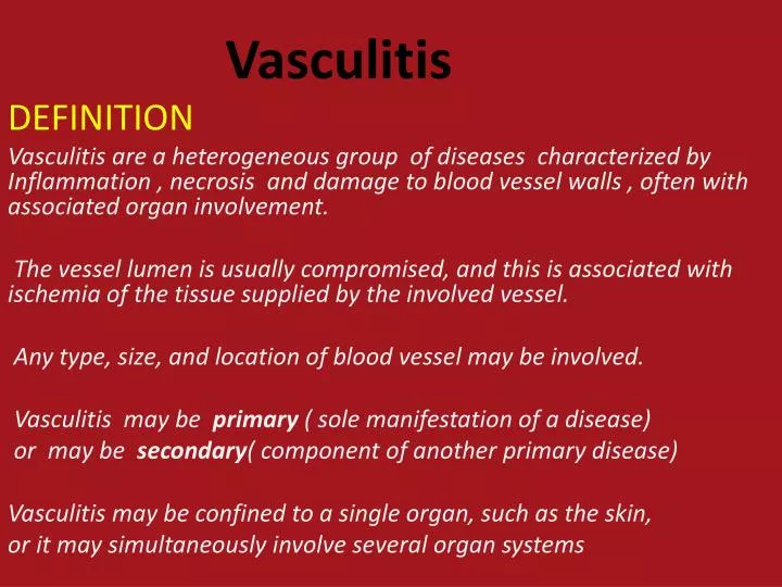
Vasculitis DEFINITION Vasculitis are a heterogeneous group of diseases characterized by Inflammation , necrosis and damage to blood vessel walls , often with associated organ involvement. The vessel lumen is usually compromised, and this is associated with ischemia of the tissue supplied by the involved vessel. Any type, size, and location of blood vessel may be involved. Vasculitis may be primary ( sole manifestation of a disease) or may be secondary( component of another primary disease) Vasculitis may be confined to a single organ, such as the skin, or it may simultaneously involve several organ systems
Classification of Vascilites Large vessel .Giant cell arteritis &/or Polymyalgiarheumatica. • Takayasu'sarteritis Medium vessel Classical polyarteritisnodosa • Kawasaki disease (in childhood) Small vessel • Microscopic polyangiitis • Wegener's granulomatosis • Churg-Strauss syndrome • Henoch-Schönleinpurpura • Mixed essential cryoglobulinaemia Othersas Behcets disease.
PATHOPHYSIOLOGY AND PATHOGENESIS of Vasculitis • Generally, most of the vasculitic syndromes are assumed to be mediated at least in part by immunopathogenic mechanisms that occur in response to certain antigenic stimuli • it is unknown why some individuals might develop vasculitis in response to certain antigenic stimuli,whereas others do not. • It is likely that a number of factors are involved in the ultimate expression of a vasculitic syndrome. These include the genetic predisposition, environmental exposures, and the regulatory mechanisms associated with immune response to certain antigens.
deposition of immune complexes in vessel walls is the most widely accepted pathogenic mechanism of vasculitis. • The actual antigen contained in the immune complex has only rarely been identified in vasculitic syndromes. • In this regard, hepatitis B antigen has been identified in some patients with systemic vasculitis, most notably in polyarteritis nodosa . • The syndrome of essential mixed cryoglobulinemiais strongly associated with hepatitis C virus infection
. The mechanisms of tissue damage in immune complex–mediated vasculitis • antigen-antibody complexes are formed and are deposited in vessel walls . • The deposition of complexes results in activation of complement components, particularly C5a, which is strongly chemotactic for neutrophils. • These cells then infiltrate the vessel wall, phagocytose the immune complexes, and release their intracytoplasmic enzymes, which damage the vessel wall. • As the process becomes subacute or chronic,mononuclear cells infiltrate the vessel wall. • This will compromise of the vessel lumen with ischemic changes in the tissues supplied by the involved vessel.
Clinical features of Vasculitis • The clinical features of vasculitis are due to a combination of local tissue ischaemia (caused by vessel inflammation and narrowing) and the systemic effects of widespread inflammation. • Systemic vasculitis should be considered in any patient with fever, weight loss, fatigue, evidence of multisystem involvement, rashes, raised inflammatory markers and abnormal urinalysis. • Early diagnosis and management are essential to prevent irreversible organ damage. • .
Vasculitis may be difficult to distinguish from widespread malignancy, occult sepsis (particularly subacute bacterial endocarditis & meningococcal septicaemia). cholesterol emboli. atrialmyxoma . antiphospholipid syndrome. • The key to recognition is the presence of multisystem involvement
Clinical Features of Vasculitis include Constitutional symptoms Fever Weight loss Fatigue purura Lividoreticularis Digital infarction Musculoskeletal Arthralgia Arthritis Cardiovascular pulselessness and ⁄or bruits common in large vessel disease Claudication Aneurysms Pulmonary Alveolar hemorrhage Nodules Gastrointestinal Bowel ischemia and ⁄or infarction Renal Glomerulonephritis Nephrotic syndrome Renovascular involvement Hypertension Neurologic Mononeuritis multiplex Visual disturbance Stroke lightheadedness Laboratory abnormalities Anemia Eosinophilia Elevated acute phase reactions Renal insufficiency Active urinary sediment
Conditions That Can Mimic Vasculitis • Infectious diseases • Bacterial endocarditis • Disseminated gonococcal infection • Pulmonary histoplasmosis • Coccidioidomycosis • Syphilis • Lyme disease • Rocky Mountain spotted fever • Coagulopathies/thrombotic microangiopathies • Antiphospholipid antibody syndrome • Thrombotic thrombocytopenic purpura
Neoplasms Emboli • Atrial myxoma cardiac emboli • Lymphoma cholestrol emboli • Carcinomatosis • Drug toxicity • Cocaine • Others • Sarcoidosis • Goodpasture’s syndrome • Amyloidosis • Migraine • Cryofibrinogenemia
Investigations in Vasculitis • If vasculitis is suspected, the diagnosis should ideally be confirmed by tissue biopsy. • Skin biopsies are easily obtained. • Nasal septal tissue can be taken from areas of ulceration or granulation. • Muscle biopsy is positive in about 50% of patients with muscle pain. . The most important bedside test is the urine dip test for protein and blood, and subsequent microscopy, since the prognosis of vasculitis is often determined by the degree of renal involvement. In patients with abnormal renal function and active urinary sediment, renal biopsy should be considered.
Visceral angiography to detect microaneurysms (e.g. classical polyarteritis nodosa) is most useful where involved tissue is not available to biopsy. • ESR usually elevated in vasculitis • CRP • C-ANCA & p-ANCA
Antineutrophilcytoplasmic antibodies (ANCA) • are directed against enzymes present in neutrophil granules. • Two main patterns of immunofluorescence are distinguished: cytoplasmic (c-ANCA) and perinuclear (p-ANCA). • c-ANCA are usually directed against proteinase 3 and are particularly associated with Wegener's granulomatosis and Churg-Strauss syndrome. • p-ANCA are usually directed against myeloperoxidase and associate with microscopic polyangiitis. • However, positive ANCAs occur in many other diseases, including malignancy, infection (bacterial and HIV), inflammatory bowel disease, RA, lupus and pulmonary fibrosis. Therefore, the diagnosis of these conditions cannot be made or refuted on the ANCA test alone.
Large Vessel Vasculitis 1- Giant cell arteritis 2- Polymyalgia rheumatica 3-Takayasu's arteritis
GIANT CELL ARTERITIS (GCA)(Temporal Arteritis) • GCA also known as temporal arteritis or cranial arteritis • is a large vessel vasculitis predominately affecting branches of the temporal and ophthalmic arteries. • The mean age of onset is 70 years . • 4:1 female:male ratio.
Clinical features of Giant cell arteritis • The onset of symptoms may be abrupt but is often insidious over the course of several weeks or months. • The most important clinical features are: 1- Headache. This is usually the first symptom and is often localised to the temporal or occipital region, with scalp tenderness. 2- Jaw pain. This is brought on by chewing or talking and is due to ischaemia of the masseters.
3-Visual disturbance. The most important complication of GCA is monocular blindness which is almost never reversible The optic nerve head is supplied by the posterior ciliary artery, vasculitis of which leads to occlusion and acute anterior ischaemic optic neuropathy. Damage to the optic nerve results in loss of visual acuity and field, reduced colour perception and pupillary defects. Sudden visual symptoms in one eye, leading rapidly to blindness, constitute the most common pattern. On fundoscopy the optic disc may appear pale and swollen with haemorrhages, but these changes may take 24-36 hours to develop. Once blindness has occurred corticosteroids have a negligible effect but are indicated to prevent blindness in the other eye.
4- There may be associated constitutional symptoms of anorexia, fatigue, weight loss, fever, depression and general malaise. 5- Occasionally presentation is with neurological complications that include transient ischaemic attacks, brain-stem infarcts and hemiparesis.
Investigations • The ESR usually elevated above 50 mm/hour in 90% of cases. ( in some cases the ESR may be normal mainly in those with acute presentationis , in the this situation the CRP may be more helpful ,& usually elevated ). • Temporal artery biopsy should be obtained. corticosteroid treatment should not be delayed whilst the biopsy is organised. Characteristic biopsy findings are fragmentation of the internal elastic lamina with necrosis of the media in combination with a mixed inflammatory cell infiltrate (lymphocytes, plasma cells and eosinophils). However, 'skip' lesions are common and a negative biopsy does not exclude the diagnosis.
Management of GCA • If GCA is suspected, systemic corticosteroid (prednisolone 60 mg daily) should be started immediately to prevent visual loss. • Steroid reduction should be guided by symptoms and ESR, aiming for approximately 10 mg daily by 6 weeks. Thereafter, doses should be reduced by 1 mg per month • Maintenance therapy is required for at least 1 year, and occasionally for the rest of the patient's life. • Relapse occurs in 30%, and is an indication to restart high-dose steroids with additional immunosuppressive agents, typically azathioprine or methotrexate
. Patients with known GCA should be advised to take 60 mg prednisolone and seek prompt medical advice should they experience any recurrence of headache or visual disturbance.
POLYMYALGIA RHEUMATICA (PMR) PMR is a clinical syndrome of muscle pain and stiffness with an increased ESR. It is not a true vasculitis but there is a close association with giant cell arteritis. The prevalence is approximately 20 per 100 000 over the age of 50. The mean age of onset is 70. Women are affected more often than men in a ratio of 3:1.
Clinical features of PMR • The cardinal features are muscle stiffness and pain, symmetrically affecting the proximal muscles of the neck, upper arms and, less commonly, the buttocks and thighs. • There is marked early morning stiffness, often with night pain. • Constitutional features of weight loss, fatigue, depression and night sweats also occur. • On examination there may be stiffness and painful restriction of active shoulder movement but passive movements are preserved. • Muscles may be tender to palpation but there should not be muscle-wasting; if there is, then primary muscle or neurological disease is more likely
CONDITIONS THAT MAY MIMIC POLYMYALGIA RHEUMATICA • Fibromyalgia • Hypothyroidism • Cervical spondylosis • Rheumatoid arthritis • Inflammatory myopathy (particularly inclusion body myositis) • Systemic vasculitis • Malignancy
Investigations in PMR • ESRis elevated above 40 mm/hour In the majority of patients • Very occasionally the ESR is low, usually in the acute situation where there has not been sufficient time for it to rise. In this situation the CRPmay be elevated prior to the ESR. • There may be a normochromic, normocyticanaemia
Management of PMR • The only effective treatment is corticosteroids. • prednisolone should be started at a dose of 15 mg daily. • The majority of patients should have a dramatic response within 72 hours. • If there is no response by 72 hours or an incomplete response by 7 days, then the diagnosis is not PMR. • If there has been a good response to prednisolone, the daily dose should be reduced to 10 mg after 4 weeks and then by 1 mg per month, assuming that symptoms remain controlled.
Most patients need steroids for an average of 12-18 months and osteoporosis prophylaxis with bisphosphonates should be considered. Some patients require steroid-sparing agents such as methotrexate or azathioprine, particularly if prednisolone cannot be withdrawn at 2 years or is needed at doses greater than 7.5 mg daily. Approximately 15-20% of patients develop features of giant cell arteritis at some point in the course of their disease.
TAKAYASU'S ARTERITIS • Takayasu's disease is a chronic inflammatory granulomatous panarteritis of elastic arteries. • The vessels most commonly involved are the aorta and its branches, and the carotid, ulnar, brachial, radial and axillary arteries. Pulmonary arteries are occasionally affected. • It is more common in women (female:male ratio 8:1) with a typical onset at the age of 25-30 years. • It has a world-wide distribution but is most common in Asia. • The aetiology is unknown.
.The usual presentation is with claudication and systemic symptoms of fever, arthralgia and weight loss. • Clinical examination may reveal loss of pulses, bruits, hypertension and aortic incompetence. • Laboratory investigations are usually non-specific, with high ESR and normocytic, normochromic anaemia. • Diagnosis is usually based on angiographic findings of coarctation, occlusion and aneurysmal dilatation.
Diagnostic Criteria for TA 1-Age less than 40 years. 2-Claudication of extremeties. 3-Decreased brachial artery pulse. 4-BP difference more than 10 mmHg between arms. 5-Bruit over subclavian arteries & aorta. 6-Arteriogram abnormalities: occlution or narrowing in aorta or main branches. Must have 3\6 criteria for diagnosis.
. The 5-year survival rate is ∼80%. Treatment • Most patients respond to initial high-dose oral prednisolone (1-2 mg/kg daily). • Additional therapy with methotrexate or cyclophosphamide is usually required. • Reconstructive vascular surgery should be avoided during periods of active inflammation but may benefit selected patients, especially those with hypertension secondary to aortic or renal lesions
Medium – Sized Arteritis 1- Classical polyarteritis nodosa. 2- Kawasaki disease.
CLASSICAL POLYARTERITIS NODOSA (PAN) • Classical PAN is a necrotising vasculitis characterised by transmural inflammation of medium-sized to small arteries. • PAN is a rare disorder with an annual incidence of 2 per million in most populations. • All age groups can be affected, with a peak incidence in the fourth and fifth decades, and a male:female ratio of 2:1. • Hepatitis B is a risk factor, and the incidence of PAN is higher in the areas, where hepatitis B infection is endemic
Clinical presentation is with myalgia, arthralgia, fever and weight loss in combination with manifestations of multisystem disease. • The most common skin lesions are palpable purpura, ulceration, infarction and livedo reticularis . • In 70% of patients arteritis of the vasa nervorum leads to neuropathy which is typically symmetrical and affects both sensory and motor function. • Severe hypertension and/or renal impairment may occur due to multiple renal infarctions. glomerulonephritis is rare .
Diagnosis is confirmed by finding multiple aneurysms and smooth narrowing of either the mesenteric, hepatic or renal systems on angiography. • Tissue biopsy may be definitive (muscle or sural nerve), even in the absence of angiographic abnormality. Treatment • Treatment for hepatitis B-related disease is to remove the source of the antigen, i.e. antiviral therapy. • Corticosteroids and cyclophosphamide are the treatment of choice for idiopathic disease. • Mortality is less than 20%, although relapse occurs in up to 50% of patients
Polyarteritis Nodosa Micro “Berry” aneurysms
Kawasaki Disease • Kawasaki disease is an acute systemic disorder of childhood that predomintely occurs in Japan( 800 cases per million in children under the age of 5 ). • The disease resembles a viral exanthem or stevens – Johnson syndrome. • Although the causative trigger is unknown, it has been associated with Mycoplasma and HIV infection in some cases. • The clinical features often develop abruptly.
Features of Kawasaki Disease* • Fever persisting > 5 days • Bilateral conjunctival congestion • Erthema of lips, buccal mucosa and tongue • Acute non-purulent cervical lymphadenopathy • Polymorphous exanthema • Erythema of palms and soles(oedema followed by desquamation) • Coronary dilatation * Five out of six clinical features, or four out of six clinical features with evidence of Coronary dilatation, are required for diagnosis.
Cardiovascular complications include myocarditis, pericarditis, coronary aneurysms, transient coronary artery dilatation, myocardial infarction due to coronary thrombosis.peripheral vascular insufficieny and gangerene. • Investigations that favour KD include : • polymorphonuclear leucocytosis, thrombocytosis, raised ESR and CRP and circulating antiendothelial cell antibodies.
Treatmentis with aspirin (5 mg/kg daily for 14 days) and intravenous gammaglobulin (400 mg/kg daily for 4 consecutive days). • Steriods should be avoided because of the risk of worsenin the coronary artery dilatation . • coronary artery changes are usually monitored weekly by two-dimensional echo for 4 weeks, by which stage most children have recovered. • The overall mortality is less than 2% • Relapse is rare , but if there is coronary artery involvement long –term follow –up is necessory.
Small Vessel Disease of Arterioles, Venules & Capillaries • Microscopic polyangiitis • Wegener's granulomatosis • Churg-Strauss syndrome • Henoch-Schönlein purpura • Mixed essential cryoglobulinaemia ANCA-ASSOCIATED VASCULITIS Microscopic polyangiitis, Wegener's granulomatosis and Churg-Strauss syndrome can be grouped together as 'ANCA-associated vasculitis',
Sites of Involvement of ANCA Vasculitis 1. Kidney - most common (75%-90%) 2. Lung - 50% 3. Skin + Kidney - 33% 4. CNS - 25%
Microscopic polyangiitis (MPA) Classic presentation is with rapidly progressive glomerulonephritis often associated with pulmonary alveolar haemorrhage. Cutaneous and gastrointestinal involvement, similar to PAN, is common. MPA is more common than PAN, with an annual incidence of 8 per million . Other features include neuropathy (15%) and pleural effusions (15%). Patients are usually p-ANCA-positive.
Wegener's granulomatosis (WG) • The annual incidence of WG is 5-10 per million. • The most common presentation is with upper airway involvement (typically epistaxis, nasal crusting and sinusitis), haemoptysis, mucosal ulceration and deafness due to serous otitis media. • Symptoms may have been present for several months and erroneously attributed to infection or allergy. • The most common ocular abnormality is proptosis, due to inflammation of the retro-orbital tissue. This may cause diplopia due to entrapment of the extraocular muscles, or loss of vision due to optic nerve compression .
Untreated nasal disease ultimately leads to destruction of bone and cartilage. • Migratory pulmonary infiltrates and nodules occur in 50% of patients. • A minority of patients present with glomerulonephritis. • Patients are usually c-ANCA-positive.