Download
1 / 27
270 likes | 406 Vues
Diffusion Tensor-Magnetic Resonance Imaging (DT-MRI) Project. Physics 30 Outcomes. This lesson will address the following outcomes from: Physics 30 Program-Of-Study Forces & Fields

E N D
Diffusion Tensor-Magnetic Resonance Imaging (DT-MRI) Project
Physics 30 Outcomes This lesson will address the following outcomes from: Physics 30 Program-Of-Study Forces & Fields General Outcomes 3: Students will explain how the properties of electric and magnetic fields are applied to numerous devices 30-B3.2sts: explain that the goal of technology is to provide solutions to practical problems and that the appropriateness, risks and benefits of technologies need to be assessed for each potential application from a variety of perspectives, including sustainability International Baccalaureate HL Physics Program-of-Study Option I – Medical Physics I.2.12: Outline the basic principles of nuclear magnetic resonance (NMR) imaging
Introduction: Brain Tumours Brain tumours can be benign or malignant as well as primary or secondary:
Benign Tumours • Benign brain tumours do not contain cancer cells: • Usually, benign tumours can be removed, and they seldom grow back. • The border or edge of a benign brain tumour can be clearly seen. Cells from benign tumours do not invade tissues around them or spread to other parts of the body. However, benign tumours can press on sensitive areas of the brain and cause serious health problems.
Malignant Tumours • Malignant brain tumours contain cancer cells: • Malignant brain tumours are generally more serious and often is life threatening. • They are likely to grow rapidly and crowd or invade the surrounding healthy brain tissue.
Primary/Secondary Tumours • Primary brain tumours: • Tumours that begin in the brain tissue • Secondary brain tumours: • Cancer that spread from its original place to another part of the body. • Secondary tumours in the brain are far more common than primary brain tumours.
Treatments of Brain Tumours • Treatment of brain tumours (gliomas): • The standard treatment is to kill or remove the glioma cells. Of course, this can only work if the surgeon or radiologist can find these cells. • Unfortunately, there are inevitably occult cancer cells, which may infiltrate several centimeters beyond the clinically apparent lesion and are not found even by today's sophisticated imaging techniques. Occult cells surrounding tumour
Treatment of Brain Tumours Due to the limited ability to detect occult glioma cells, clinicians currently add a uniform margin of 2cm or more beyond the visible abnormality, and irradiate that volume. However, researchers believe expanding the tumour 2cm in every direction is killing more cells than necessary and that some hidden cells may grow outside the 2cm envelope.
Treatment of Brain Tumours Evidence, however, suggests that glioma growth is not uniform - growth is favored in certain directions and impeded in others. This means it is important to determine, for each patient, which areas are at high risk of harboring occult cells. Glioma has squished this area of brain to be ‘abnormal’, during scans they are also classified as unhealthy cells Occult cells growing towards the back of the brain
What is a DT-MRI? Diffusion Tensor Magnetic Resonance Imaging (DT-MRI) and Machine Learning tries to predict the location of these occult cells by learning the growth patterns exhibited by gliomas in previous patients. These new images allow researchers to detect direction and rate of water flow
What is a DT-MRI? Diffusion Tensor Magnetic Resonance Imaging (DT-MRI) is a new technique in MRI that scans the fibrous muscle structure of the brain to determine the rate of diffusion and a preferred direction of diffusion of water. It has been noted that occult cancer cells tend to grow with these preferred rate and direction of diffusion of water
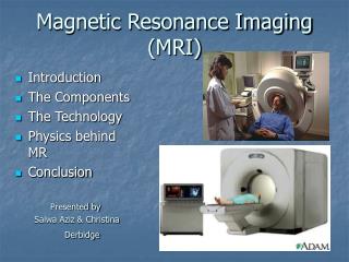
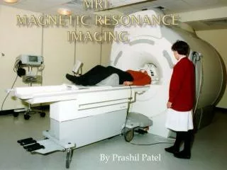
How does MRIs work? MRI Magnetic Resonance Imaging (MRI) scanners contain magnets. The magnetic field produced by an MRI is about 10 thousand times greater than the earth's. MRI is based on the way certain atomic nuclei respond to radio waves while in the presence of a magnetic field.
How does MRIs work? The magnetic field forces hydrogen atoms in the body to line up in a certain way (similar to how the needle on a compass moves when you hold it near a magnet). Hydrogen atoms are ideal to work with because they only have one proton and have a large magnetic moment, meaning these atoms will line up in the direction of a magnetic field. The hydrogen atoms in our bodies spin in all sorts of directions.
How does MRIs work? However, when a patient is inside the MRI, the hydrogen protons will line up either towards the feet or the head. Since the protons an align in two directions, most will be cancelled out, but one or two out of each million will not be cancelled out and hence create a net magnetic force. One or two in each millions is not a lot, but there are about 4.7x1027 hydrogen atoms in each human body
How does MRIs work? When radio waves are sent towards the one or two (in each million) unpaired hydrogen atom, the proton absorbs the energy needed to create a spin. This spin gives off a particular frequency in a specific direction. This frequency is known as the Larmour frequency. Different types of tissues send back different signals/frequencies. For example, healthy tissue sends back a slightly different signal than cancerous tissue.
How does MRIs work? Now that we have the signals, the machine will convert these frequencies into a picture. MRIs take pictures of 3-D bodies, but the images in 2-D of MRIs are in 2-D. So how can we create the 3-D image using these 2-D scans?
How does MRIs work? Think of a loaf of bread, we can slice it up and look at each piece individually. MRIs also create 2-D images are called slices. The images can be stacked to create the 3-D image that was scanned.
How does MRIs work? Each column, T1, T1c, and T2 scan different aspects of the brain T1 weighted: An MRI that highlights fat locations. T1c weighted: An MRI that is taken after the injection of the contrast agent gadolinium, a contrast agent that can make abnormalities such as tumours clearer due to the element's special magnetic properties. T2 weighted: An MRI that highlights water locations.
How does DT-MRI help? With the new technique of DT-MRI provides more data for researchers to learn the where occult are and predict the direction of growth of the glioma cells.
What is Machine Learning? Machine Learning is a scientific discipline concerned with designing and developing programs that allow computers to learn based on data (“thinking” computers). A program is created to find gliomas and autonomously find the tumour region within a brain image which may contain occult cancer cells.
What are the advantages? • Result of this Medical and Computer Science collaboration: • Advanced imaging techniques help us better characterise gliomas in the future • Create an image-based database to allow machine learning analysis of all the clinically available data • Through machine learning analysis, develop computer algorithms to allow us to automate tumour segmentation, predict tumour behaviour and predict location of clinically occult glioma cells
Questions about MRIs? MRIs are proving to be an extremely useful technique for imaging blood flow and soft tissue in the body. It is the preferred diagnostic imaging technique for studying the brain and the central nervous system 1. Describe the basic principles employed to collect an MRI scan of body tissues. 2. What properties of the hydrogen atom makes it such a useful atom for MRI diagnosis?
Questions about MRIs? 3. Give two diagnostic applications that MRI scans are used for. 4. Discuss the advantages and disadvantages that a MRI scan has when compared to other diagnostic techniques. 5. If the radio waves used to excite the hydrogen atoms are 2.45x10⁴ Hz, what is the corresponding wavelength?
Questions about MRIs? 6. A magnetic resonance imaging (MRI) machine uses an enormous and extremely strong magnet to study a patient's body. The magnet, which has its north pole at the patient's head and its south pole at the patient's feet, is actually a coil of superconducting wire through which electric charges flow. This fancy electric system seems unnecessary; why can't the technicians simply put a large number of north magnetic poles near the patient's head and an equal number of south magnetic poles near the patient's feet?
Questions about MRIs? 7. Aluminum isn't normally magnetic, but as you carry a large aluminum tray toward the magnet, you find that the magnet repels the aluminum. Explain. 8. You eventually manage to get the aluminum tray up to the magnet. As long as the tray doesn't move, it experiences no magnetic forces. But when you drop it, it falls past the magnet remarkably slowly. What slows down its fall?
What is an MRI? Resources: http://clinicaltrials.gov/ct2/show/NCT00330109 http://www.medicinenet.com/brain_tumor/page2.htm http://www.physics247.com/physics-help/mri.shtml http://www.phys.unsw.edu.au/~jw/FAQ.html#scan http://healthguide.howstuffworks.com/mri-dictionary.htm http://healthguide.howstuffworks.com/mri-dictionary.htm http://rabi.phys.virginia.edu/1060/1999/PS3a.html http://education.jlab.org/qa/mathatom_04.html
Centre for Mathematics Science and Technology Education (CMASTE) 382 Education South University of Alberta Edmonton AB T6G 2G5 www.CMASTE.ca To download: select Outreach, Alberta Ingenuity Resources and Centre for Machine Learning Filename: AICML6BrainTumourAnalysis Centre for Machine Learning Department of Computing Science University of Alberta 2-21 Athabasca Hall Edmonton AB T6G 2E8 (780) 492-4828 www.machinelearningcentre.ca Alberta Ingenuity 2410 Manulife Place, 10180-101 Street Edmonton AB T5J 3S4 (780) 423-5735 www.albertaingenuity.ca