Download
1 / 40
420 likes | 828 Vues
Advanced Biochemistry for Biotechnology,. Electron Transfer Chain. Electron Transfer. An electron transfer reaction: A ox + B red A red + B ox A ox is the oxidized form of A (the oxidant) B red is the reduced form of B (the reductant).

E N D
Advanced Biochemistry for Biotechnology, Electron Transfer Chain
Electron Transfer An electron transfer reaction: Aox + Bred Ared + Box Aox is the oxidized form of A (the oxidant) Bred is the reduced form of B (the reductant). For such an electron transfer, one may consider two half-cell reactions: Aox + n e- Ared e.g., Fe+++ + e- Fe++ Box + n e- Bred
Aox + n e- Ared Box + n e- Bred For each half reaction: E = E°'– RT/nF (ln [reduced]/[oxidized]) e.g., for the first half reaction: E = E°' – RT/nF(ln[Ared]/[Aox]) E = voltage, R = gas const., F = Faraday, n = # of e-. When [Ared] = [Aox], E = E°'. E°' is the mid-point potential, or standard redox potential, the potential at which [oxidant] = [reductant] for the half reaction.
For an electron transfer: DE°' =E°'(oxidant) – E°'(reductant) = E°'(acceptor) – E°'(donor) DGo' = – nFDE°' (E°' is the mid-point potential) An electron transfer reaction is spontaneous (negative DG) if E°' of the donor is more negative than E°' of the acceptor, i.e., when there is a positive DE°'.
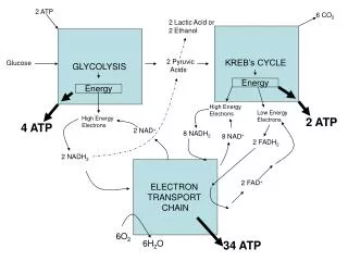
Consider transfer of 2 electrons from NADH to oxygen: a. ½ O2 + 2H+ + 2e- H2O E°' = +0.815 V b. NAD+ + 2H+ + 2e- NADH + H+ E°' = -0.315 V Subtracting reaction b from a: c. ½ O2 + NADH + H+ H2O + NAD+ DE°'= +1.13 V DG = - nFDEo' = – 2(96,494)(1.13) = – 218 kJ/mol
ElectronCarriers NAD+/NADH and FAD/FADH2 were introduced earlier. FMN (Flavin MonoNucleotide) is a prosthetic group of some flavoproteins. It is similar in structure to FAD (Flavin Adenine Dinucleotide), but lacking the adenine nucleotide. FMN (like FAD) can accept 2 e- + 2 H+ to form FMNH2.
FMN, when bound at the active site of some enzymes, can accept 1 e- to form the half-reduced semiquinone radical. The semiquinone can accept a 2nd e- to yield FMNH2. Since it can accept/donate 1 or 2 e-, FMN has an important role mediating e- transfer between carriers that transfer 2e- (e.g., NADH) & those that can accept only 1e- (e.g., Fe+++).
Coenzyme Q (CoQ, Q, ubiquinone) is very hydrophobic.It dissolves in the hydrocarbon core of a membrane. It includes a long isoprenoid tail, with multiple units having a carbon skeleton comparable to that of isoprene. In human cells, most often n = 10. Q10’s isoprenoid tail is longer than the width of a bilayer.It may be folded to yield a more compact structure, & is postulated to reside in the central domain of a membrane, between the 2 lipid monolayers.
The quinone ring of coenzyme Q can be reduced to the quinol in a 2e- reaction: Q + 2 e- + 2 H+ QH2.
When bound to special sites in respiratory complexes, CoQ can accept 1e-to form a semiquinone radical (Q·-). Thus CoQ, like FMN, can mediate between 1e- & 2e- donors/acceptors.
Coenzyme Q functions as a mobile e- carrier within the mitochondrial inner membrane. Its role in trans-membraneH+ transport coupled to e- transfer (Q Cycle) will be discussed later.
Heme is a prosthetic group of cytochromes. Heme contains an iron atom in a porphyrin ring system. The Fe is bonded to 4 N atoms of the porphyrin ring.
Hemes in the 3 classes of cytochrome (a, b, c) differ slightly in substituents on the porphyrin ring system. A common feature is 2 propionate side-chains. Only heme c is covalently linked to the protein via thioether bonds to cysteine residues.
Heme a is unique in having a long farnesyl side-chain that includes 3 isoprenoid units.
In the RasMol ( a molecular graphics program intended for the visualisation of proteins, nucleic acids and small molecules)display of heme c at right, the porphyrin ring system is displayed as ball & sticks, while Fe is displayed as spacefill. The heme iron can undergo a 1e- transition between ferric and ferrous states: Fe+++ + e- Fe++
The porphyrin ring is planar. The heme Fe is usually bonded to 2 axial ligands, above & below the heme plane (X,Y) in addition to 4 N of porphyrin. Axial ligands may be S or N atoms of amino acid side-chains. Axial ligands in cyt c are Met S (yellow) and His N (blue). A heme that binds O2 may have an open (empty) axial ligand position.
Cytochromesare proteins with heme prosthetic groups. They absorb light at characteristic wavelengths. Absorbance changes upon oxidation/reduction of the heme iron provide a basis for monitoring heme redox state. • Some cytochromes are part of large integral membranecomplexes, each consisting of several polypeptides & including multiple electron carriers. Individual heme prosthetic groups may be separately designated as cytochromes, even if in the same protein. E.g., hemes a & a3 that are part of the respiratory chain complex IV are often referred to as cytochromes a & a3. • Cytochrome c is instead a small, water-soluble protein with a single heme group.
Positively charged lysine residues (in magenta) surround the heme crevice on the surface of cytochrome c. These may interact with anionic residues on membrane complexes to which cyt c binds, when receiving or donating an e-.
Fe-S spacefill; cysteine ball & stick. Fe orange; S yellow. PDB 2FUG Iron-sulfur centers (Fe-S) are prosthetic groups containing 2,3,4 or 8 iron atoms complexed to elemental & cysteine S. 4-Fe centers have a tetrahedral structure, with Fe & S atoms alternating as vertices of a cube. Cysteine residues provide S ligands to the iron, while also holding these prosthetic groups in place within the protein.
Electron transfer proteins may contain multiple Fe-S centers. Iron-sulfur centers transfer only oneelectron, even if they contain two or more iron atoms, because of the close proximity of the iron atoms. E.g., a 4-Fe center might cycle between redox states: Fe+++3,Fe++1(oxidized)+1 e-Fe+++2, Fe++2(reduced)
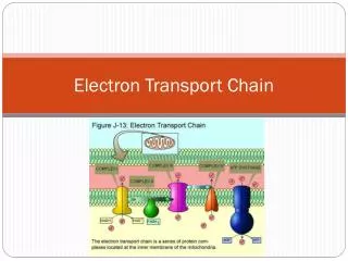
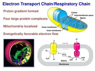
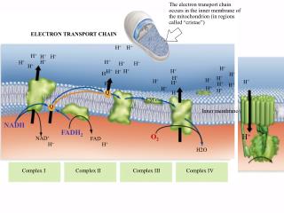
Most constitutents of the respiratory chain are embedded in the inner mitochondrial membrane (or in the cytoplasmic membrane of aerobic bacteria). The inner mitochondrial membrane has infoldings called cristae that increase the membrane area. Respiratory Chain:
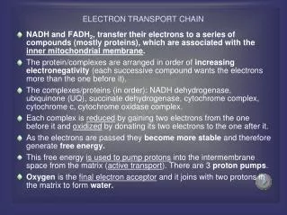
Electrons are transferred from NADH O2 via multisubunit inner membranecomplexes I, III & IV, plusCoQ&cyt c. Within each complex, electrons pass sequentially through a series of electron carriers. CoQ is located in the lipid core of the membrane. There are also binding sites for CoQ within protein complexes with which it interacts. Cytochrome c resides in the intermembrane space. It alternately binds to complex III or IV during e- transfer.
Individual respiratory chain complexes have been isolated and their composition determined. There is also evidence for the existence of stable supramolecular aggregates containing multiple complexes. E.g., complex I, which transfers electrons to coenzyme Q, may associate with complex III, which reoxidizes the reduced coenzyme Q, to provide a pathway for direct transfer of coenzyme Q between them.
Complex Name No. of Proteins Prosthetic Groups Complex I NADH Dehydrogenase 46 FMN, 9 Fe-S cntrs. Complex II Succinate-CoQ Reductase 5 FAD, cyt b560, 3 Fe-S cntrs. Complex III CoQ-cyt c Reductase 11 cyt bH, cyt bL, cyt c1, Fe-SRieske Complex IV Cytochrome Oxidase 13 cyt a, cyt a3, CuA, CuB Composition of Respiratory Chain Complexes
Mid-point potentials of constituent e- carriers are consistent with the e- transfers shown being spontaneous. Respiratory chain inhibitors include: Rotenone (a rat poison) blocks complex I. Antimycin A blocks electron transfer in complex III. CN- & CO inhibit complex IV. Inhibition at any of these sites will block e- transfer from NADH to O2.
Complex I catalyzes oxidation of NADH, with reduction of coenzyme Q: NADH + H+ + Q NAD+ + QH2 Transmembrane H+ flux associated with this reaction will be discussed in the section on oxidative phosphorylation. An atomic-level structure is not yet available for the entire complex I, which in mammals includes at least 46 proteins, along with prosthetic groups FMN & several Fe-S centers.
Complex I is L-shaped. The peripheral domain, containing the FMN that accepts 2e- from NADH, protrudes into the mitochondrial matrix. Iron-sulfurcenters are also located in the hydrophilic peripheral domain, where they form a pathway for e- transfer from FMN to coenzyme Q. A binding site for coenzyme Q is thought be close to the interface between peripheral and intra-membrane domains.
The initial electron transfers are: NADH + H+ + FMNNAD+ + FMNH2 FMNH2 + (Fe-S)oxFMNH· + (Fe-S)red + H+ After Fe-S is reoxidized by transfer of the electron to the next iron-sulfur center in the pathway: FMNH· + (Fe-S)oxFMN + (Fe-S)red + H+ Electrons pass through a series of iron-sulfur centers, and are eventually transferred to coenzyme Q. Coenzyme Q accepts 2e- and picks up 2H+ to yield the fully reduced QH2.
An X-ray structure has been determined for the hydrophilic peripheral domain of a bacterial complex I This bacterial complex I contains fewer proteins than the mammalian complex I, but includes the central subunits found in all prokaryotic & eukaryotic versions of complex I. The prosthetic groups are found to be all in the peripheral domain, that in the mammalian complex would protrude into the mitochondrial matrix.
Iron-sulfur centers are arranged as a wire, providing a pathway for e- transfer from FMN through the protein. N2, the last Fe-S center in the chain, passes e- one at a time to the mobile lipid redox carrier coenzyme Q. A proposed binding site for CoQ is close to N2 at the interface of peripheral & membrane domains.
P. L. Dutton and coworkers have called attention to the relevance of conserved distances between redox carriers within respiratory chain complexes with regard to the energy barrier at each step for electron tunneling through the protein. They have modeled electron transfers through the respiratory chain complexes, and provide an animation of the time course of electron transfer through Complex I. For more diagrams see • A review by U. Brandt (requires Annual Reviews subscription). • The Complex I Home Page
Succinate Dehydrogenase of the Krebs Cycle is also called complex II or Succinate-CoQ Reductase. FAD is the initial e- acceptor. FAD is reduced to FADH2 during oxidation of succinate to fumarate. FADH2 is then reoxidized by transfer of electrons through a series of 3 iron-sulfur centers to CoQ, yielding QH2. The QH2 product may be reoxidized via complex III, providing a pathway for transfer of electrons from succinate into the respiratory chain.
X-ray crystallographic analysis of E. coli complex II indicates a linear arrangement of electron carriers within complex II, consistent with the predicted sequence of electron transfers: FADFeScenter 1FeScenter 2FeScenter 3CoQ In this crystal structure oxaloacetate (OAA) is bound in place of succinate.
Complex IIIaccepts electrons from coenzymeQH2 that is generated by electron transfer in complexes I & II. The structure and roles of complex III are discussed in the class on oxidative phosphorylation. Cytochrome c1, a prosthetic group within complex III, reduces cytochrome c, which is the electron donor to complex IV.
Cytochrome oxidase(complex IV) carries out the following irreversible reaction: O2 + 4H+ + 4e- 2H2O The four electrons are transferred into the complex one at a time from cytochrome c.
Intramembrane domains of cytochrome oxidase (complex IV) consist mainly of transmembrane a-helices.
Metal centers of cytochrome oxidase (complex IV): heme a & heme a3, CuA (2 adjacent Cu atoms) & CuB. O2 reacts at a binuclear center consisting ofheme a3 andCuB.
Metal center ligands in complex IV: Heme axial ligands are His N atoms. Heme a is held in place between 2 transmembrane a-helices by its axial His ligands.
Heme a3, which sits adjacent to CuB, has only one axial ligand. Cu ligands consist of His N, & in the case of CuA also Cys S, Met S, & a Glu backbone O. Electrons enter complex IV one at a time from cyt c to CuA. They then pass via heme a to the binuclear center where the chemical reaction takes place. Electron transfers: cyt c → CuA→ heme a → heme a3/CuB O2 binds at the open axial ligand position of heme a3, adjacent to CuB.
O2 + 4H+ + 4e- 2 H2O Details of the reaction sequence are still debated. A Tyr-His complex adjacent to the binuclear center is postulated to have a role in O-O bond splitting. The open axial ligand position makes heme a3 susceptible to binding each of the following inhibitors: CN-, CO, and the radical signal molecule ·NO. ·NO may regulate cell respiration through its inhibitory effect, & can induce a condition comparable to hypoxia.