Download
1 / 70
750 likes | 1.16k Vues
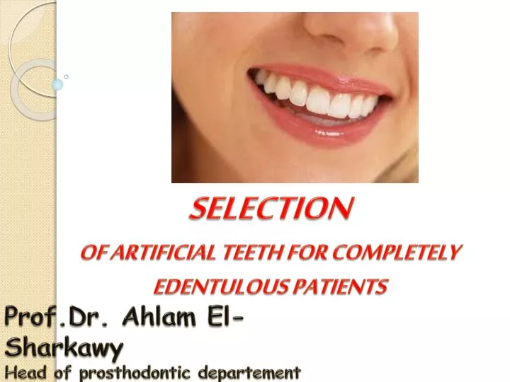
SELECTION OF ARTIFICIAL TEETH FOR COMPLETELY EDENTULOUS PATIENTS. Prof.Dr. Ahlam El- Sharkawy Head of prosthodontic departement Pharos University in Alexandria. Introduction.

E N D
SELECTION OF ARTIFICIAL TEETH FOR COMPLETELY EDENTULOUS PATIENTS Prof.Dr. Ahlam El-Sharkawy Head of prosthodonticdepartement Pharos University in Alexandria
Introduction The anterior teeth are primarily selected to satisfy the estheticrequirement , where as the posterior teeth are primarily selected to satisfy the functional requirements . The anterior teeth are composed of six maxillary and six mandibular teeth.
OBJECTIVES FOR TOOTH SELECTION TO CONSTRUCT THE COMPLETE DENTURE THAT………… 1. FUNCTION WELL EFFICIENT FOR MASTICATION. 2. ALLOW PATIENT TO SPEAK NORMALLY. 3. ESTHETICALLY PLEASING. 4. PRESERVE TISSUE STRUCTURES OF THE GNATHODYNAMIC SYSTEM 5. SHOULD MAINTAIN THE VERTICAL DIMENSION
Anterior teeth selection Guides for the anterior teeth selection • Pre-extraction guides • Study cast • Photographs • Radiographs • Extracted teeth • Old denture.
If the pre-extraction records are not available systematic procedure can be followed to select the teeth which will harmonize with the patient’s facial characteristics
Maxillary anterior teethSelection • Size • Shape (Form) • Shade (color).
Selection of size • Width • Length.
Width of the anterior teeth Width of the anterior teeth
a. Bizygomatic distance • The average width of the maxillary central incisor is estimated to be 1:16 of the bizygomatic width • Bizygomatic widththe distance between the cheek bones measured just in front of the ears.
a. Bizygomatic distance • The combined width of the six maxillary anteriors is about one third the bizygomatic width.
b. The width of the nose • An estimation of the position of cusp tip of the upper natural canine can be found by extending parallel lines from the lateral surface of the ala of the nose onto the labial surface of the upper occlusion rim.
c. Corners of the mouth • The distance measured between the two commisures (angles of the mouth ) will represent the width of the upper six anteriors from the distal surface of the canine to the distal surface of the other canine.
Corners of the Mouth • Mark corners at rest (#7 wax spatula) • Position of distal of the canines
Length • The necks of the upper anterior teeth overlap the anterior ridge by 2-3 mm cervically, and the incisive edges of the centrals must show below the relaxed lip by 2-3 mm in a young person and less than half that amount in an elderly patient.
Amount of tooth shownAbout 2 mm of teeth are shown when lip is relaxed.
High Lip Line • Highest point of upper lip when smiling • Cervical necks lie at or above this line • If shorter teeth are selected, esthetics compromised
Guidelines for Setting Anterior Teeth High Lip Line Corner of Mouth Angulation is as important as midline
FORM OF THE ANTERIOR TEETH . • The guides for selecting the form of anterior teeth : • 1- Shape of the face. • 2- Shape of the arch.
1-The shape of the face . • According to Leon Williams classification the human face has been classified into • Three types • Square • Tapering • Ovoid
The shape of the maxillary central incisor coincides with the inverted form of the face
Selection of the form • Shape of the face. It was claimed that the shape of the upper central incisor bears a definite relationship to the shape of the face. Thus if one of these teeth is enlarged and the incisal edge placed above the brows with the neck of the tooth on the chin, then the outline of the tooth would nearly coincide with that of the face.
SQUARE • Facial form - the width of the forehead, zygomatic arch and mandibular angle is equal. • Profile - straight and flat ala area • Incisal form - mesial and distal lines are almost parallel till the length of 2/3rd from incisal edge.
ovoid • Facial form - width of zygomatic arch is wider than the distance of forehead. • Profile - ovoid ala area • Incisal form - mesial and distal lines are curved.
Tapering • Facial form - the width becomes narrow from forehead towards the zygomatic arch and mandibular angle. • Profile - curved or flat • Incisal form - mesial and distal lines become narrow from incisal edge to cervical end.
Selection of the form • 2-Shape of the arch.
Patient’s sex and age: • A cuboidal tooth form is required for the males • A spheroidal form for the females. • Teeth for males should have a less convexity than for females. • The incisal edge of older patient is flat to resemble attrition of natural teeth.
Dentogenic concept (SPA) • Sex, personality, age are the factors which will determine the form of the anterior teeth
Personality • The dentist should select and arrange teeth so that it improves the patient’s personality. The patient can be vigorousoredelicat. • More squarish and large teeth are selected for vigorous people
MUSCULINE Prominent teeth Large sized teeth Square arch form Square labial surface Square incisal edges and corners Darker shade Flat smiling line Canine – cervical area is more visible and prominent Feminine Smaller teeth Round corners Curved contours & arch form Curved labial surface Round incisal edges and corners Delicate appearance Curved smiling line Canine – mesial 3rd only seen
Younger people Lighter shade More incisal translucency Minimal wear of incisal edge Curved smiling line Pointed canines Older people Darker shade Less incisal translucency increased wear of incisal edges Flat smiling line Loss of tips of canine Age
Check Shade Against the Patients Face • Check shade of existing denture • Ask if patient wants same shade or change • Allow patient to view shade against lip with mirror • Obtain patient approval
Patients age – With age, darker, while lighter teeth are suitable for young patients. Patients complexion—light teeth for fair skin, blue eyes, dark teeth usually for dark skin and black eyes.
The anterior teeth from the lightest to the darkest • Upper central incisor • The upper lateral • The lower central and lateral • The upper and lower canines are the darker.
1-Size of posterior denture teeth a-Length: This is determined by the amount of denture space available between the upper and lower ridges.
b-Width From distal of canine to the tuberosity in the maxillary denture, and to the retromolar periphery of the lower denture. The teeth with a narrower buccolingual width than the natural teeth are selected.
Posterior Tooth Selection • Buccolingual size can affect the tongue space • smaller teeth if tongue space is limited
Posterior teeth selection Posterior teeth should have a small bucco-lingual width to keep forces on the supporting structure to a minimum. The mesiodistal measurements of the upper posterior teeth is taken from the distal surface of the canine to the prominence of the tuberosity. The lower posterior teeth should not extend posterior to the mesial border of the retromolar pad.
2-Form of the occlusal surface (Mould or shape) • Cusp angle :the angle made by the slope of the cusp with the cusp plane
Cusp Height : The perpendicular distance between the tip of the cusp and its base plane
30° 20° 10° Anatomic Teeth (10°, 20°, 30°, 40°)
Cusped Teeth(Anatomic teeth) have 30-35 cusp angleIndicated when there is a well defined ridge and normal jaw relations.
Advantages of anatomic teeth 1. Esthetically acceptable 2. More efficient in cutting of food, thereby reducing forces that are directed to the supporting structures during masticatory movement. 3. They can be arranged in balances occlusion
Disadvantages of anatomic teeth 1. It is mandatory to use an adjustable articulator. 2. Eccentric records must be done for articulator adjustments 3. Clinical remount is essential to adjust the occlusion after denture settling. 4. Balanced occlusion lost when settling occurs. 5. More horizontal forces during functions. 6. Frequent relining, Fast bone resorption
Flat or Inverted Cusped TeethHave zero cusp angle with grooves to aid mastication
Non-Anatomic Teeth(0°, Rational, Monoline, etc.) • Jaw size discrepancies (Class III) • Severe ridge resorption • Poorer esthetics, due to lack of cuspal inclines