Download
1 / 49
490 likes | 496 Vues
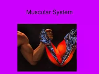
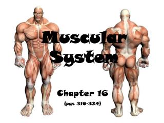
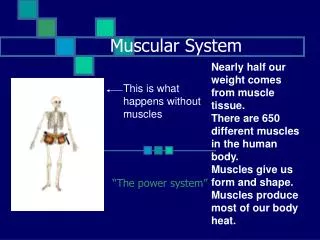
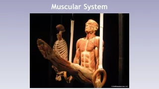
Muscular System. Muscles in Humans. There are approximately 650 muscles in the human body, each performing a unique role. Muscles are the driving force behind the musculoskeletal systems, enabling us to sit, stand, walk, talk and do anything else that requires movement.

E N D
Muscles in Humans • There are approximately 650 muscles in the human body, each performing a unique role. • Muscles are the driving force behind the musculoskeletal systems, enabling us to sit, stand, walk, talk and do anything else that requires movement. • They also keep our heart beating and help us breathe. • In order to function, they must be able to react to stimuli, contract, extend and return to their normal shape.
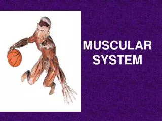
Muscular System • Muscles are responsible for all types of body movement • 3 basic muscle types are found in the body • Skeletal muscle • Cardiac muscle • Smooth muscle
Characteristics of Muscles • Muscle cells are elongated (muscle cell = muscle fiber) • Contraction of muscles is due to the movement of microfilaments • All muscles share some terminology • Prefix myo refers to muscle • Prefix mys refers to muscle • Prefix sarco refers to flesh
Nervous system to muscles • Within the nervous system, nerve impulses travel from neuron to neuron along complex nerve pathways. The junction between the parts of two such neurons is called a "synapse.“ • If a sufficient amount of the substance (called "neurotransmitter") is released, the membrane is stimulated, and a nerve impulse is triggered
Neuromuscular junction occurs when a nerve cell contacts a muscle
Myofilaments • Muscle cells are unique because of the contractible protein fibers, called myofilaments, within them. • the filaments of myofibrils constructed from proteins are of two types, thick and thin. • Thin filaments consist primarily of the protein actin. • Thick filaments consist primarily of the protein myosin. • Muscle contraction occurs because of the myosin filaments pull on actin filaments
Skeletal Muscle Characteristics • Most attach to bones by tendon • Cells are multinucleate • Striated—have visible binding • Voluntary • Cells surrounded & bundled by connective tissue
Smooth Muscle Characteristics • Has no striations • Spindle-shaped cells • Single Nucleus • Involuntary—no conscious control • Found mainly in the walls of hollow organs
Characteristics of Cardiac Muscle • Has striations • Usually has a single nucleus • Joined to another cardiac muscle cell • Involuntary • Found only in the heart • Connects cytoplasm between 2 cells (Gap junction) Gap junctions are responsible for electrochemical and metabolic coupling. They allow Each heart muscle cell is coupled to its neighbors electrically by tiny holes, producing depolarization of the heart muscle
Functions of Skeletal Muscle Produce Movement Maintain posture Stabilize joints Generate Heat Sites of Muscle Attachment Bones Cartilage Connective tissue coverings Muscle Fibers blend into a connective tissue attachment Tendon—cordlike structure Aponeurosis—sheet-like structure Properties of Muscle Irritability – ability to receive and respond to a stimulus Contractibility – ability to shorten when an adequate stimulus is received Extensibility – ability to lengthen when an adequate stimulus is received Elasticity – ability to return to normal shape Skeletal Muscle
Anatomy of a Muscle Cell • Each fiber is built up from smaller strands called myofibrils, and each myofibril contains interlaced filaments of muscle proteins.
Direction of Muscle Fibers • Relative to the Midline • RECTUS = parallel to the midline • RectusAbdominus • TRANSVERSE = perpendicular to midline • TransverseAbdominus • OBLIQUE = diagonal to midline • External Oblique
Location • Structure near which muscle is found • FRONTALIS = near FRONTAL bone • OCCIPITALIS = near OCCIPITAL bone
Size • Relative Size of Muscle • MAXIMUS = largest • Gluteus Maximus • MEDIUS = middle • Gluteus Medius • MINIMUS = smallest • Gluteus Minimus • LONGUS = longest • Fibularis Longus • BREVIS = short • Fibularis Brevis • TERTIUS = shortest • Fibularis Tertius
Number of Origins • Number of tendons of origin • BICEPS = Two • Biceps Brachii • Biceps Femoris • TRICEPS = Three • Triceps Brachii • QUADRICEPS = Four • Quadriceps Femoris
Shape • Relative Shape of the Muscle • DELTOID = triangular shape Δ • TRAPEZIUS = trapezoid shape SERRATUS = saw-toothed ♒ • RHOMBOIDEUS = rhomboid shape • TERES = round ○
Origin & Insertion • Origin – attachment to an immoveable bone • Insertion – attachment to a movable bone • ILIOCOSTALIS= attaches to the ilium & ribs (costal = ribs)
Types of Muscle--Actions • Prime mover (Agonist) – muscle with the major responsibility for a certain movement • Antagonist – muscle that opposes or reverses a prime mover • Synergist – muscle that aids a prime mover in a movement and helps prevent rotation • Fixator – stabilizes the origin of a prime mover
Frontalis: elevate eyebrows Orbicularis Oculi: close eyelid Zygomaticus: draw angle of lip upward Buccinator: draws cheeks against teeth Orbicularis Oris: closes mouth Platysma: draws lower lip down & back Cranial Aponeurosis: connects frontalis to occipitalis Temporalis: elevates mandible Occipitalis: draws scalp back Masseter: elevates mandible Sternocleidomastoid: Flexes head Draws head toward shoulder Head & Neck Muscles
Muscles of Mastication • Masseter: elevates mandible • Temporalis: elevates mandible • Medial pterygoid: elevates mandible • Lateral pterygoid: depresses mandible
Smiling Muscles Orbicularis Oculi Nasalis Levator Labii Superioris Levator Anguli Superioris Zygomaticus Risorius Frowning Muscles Frontalis Orbicularis Oris Depressor Anguli Oris Depressor Labii Inferioris Mentalis Platysma Key Muscles of Facial Expression
Intrinsic Muscles Erector Spinae: maintain posture of back/extension Spinalis Longissimus Iliocostalis Oblique Muscles: rotation of the vertebrae Semispinalis Multifidus Rotatores Muscles of Quiet Respiration Diaphragm External Intercostals Internal Intercostals—deep breaths Abdominal Muscles External Obliques Internal Obliques Transverse Abdominus Rectus Abdominus Quadratus Lumborum Muscles of the Axial Skeleton
Muscles of Scapular Stabilization • Trapezius: • Retraction (M) • Elevation (S) • Depression (I) • Upward Rotation (S, M) • Rhomboid—retraction • Levator Scapular—Elevation • Pectoralis Major—Protraction • Serratus Anterior—Protraction
Anterior Muscles of Shoulder • Deltoid • Flexion (A, M)/Extension (P, M) • Abduction (M)/Adduction (A) • Internal (A) /External Rotation (P) • Pectoralis Major • Adduction • Flexion • Extension • Internal Rotation • Biceps Brachii—Flexion
Posterior Muscles of Shoulder • Teres Major • Adduction • Extension • Internal Rotation • Latissimus Dorsi • Adduction • Extension • Internal Rotation • Triceps Brachii • Adduction • Extension
Rotator Cuff Muscles (SITS) • Supraspinatus • Abduction • Infraspinatus • External Rotation • Teres Minor • External Rotation • Subscapularis • Internal Rotation
Muscles of the Elbow/Forearm • Triceps Brachii—Extension • Bicep Brachii— • Flexion • Supination • Brachialis—Flexion • Brachioradialis— • Flexion • Pronation • Pronator Teres • Pronator Quadratus • Supinator Longus
Muscles of the Wrist & Hand • Flexor Carpi Ulnaris • Flexor Carpi Radialis • Flexor Digitorum • Extensor Carpi Ulnaris • Extensor Carpi Radialis • Extensor Digitorum Anterior (Palmar) View Posterior (Dorsal) View
Medial/Adductor Muscles: Adductor Magnus Adductor Longus Adductor Brevis Gracilis Anterior Muscles Iliopsoas—Flexion Pectineus— Flexion Adduction Sartorius— Flexion Lateral Rotation Muscles of Hip:Anterior Muscles
Muscles of Hip: Gluteal Muscles • Gluteus Maximus—Extension • Gluteus Medius—Abduction • Gluteus Minimus—Abduction • Tensor Fasciae Latae— • Flexion • Abduction ** Gluteus Minimus is under the Gluteus Medius
Muscles of Anterior Thigh • “Quadriceps” • Rectus Femoris— • Hip flexion • Knee extension • Vastus Lateralis—knee extension • Vastus Medialis—knee extension • Vastus Intermedius—knee extension • Sartorius— • Hip & Knee Flexion • Lateral Hip Rotation **Vastus Intermedius is beneath Rectus Femoris
Muscles of Posterior Thigh • “Hamstrings” • Responsible for Knee Flexion & Hip Extension • Semimembranosus • Semitendinosus • Biceps Femoris • Gastrocnemius • Knee Flexion
Muscles of the Lower Leg • Anterior Compartment • Tibialis Anterior—Dorsiflexion & inversion • Extensor Digitorum Longus • Fibularis Tertius—dorsiflexion & eversion • Posterior Compartment • Gastrocnemius—plantarflexion, knee flexion • Soleus—plantarflexion • Lateral Compartment • Fibularis Longus—plantarflexion & eversion • Fibularis Brevis—plantarflexion & eversion
Tendinitis • Inflammation of tendons and of tendon-muscle attachments • Many patients report stressful situations in their life in correlation with the beginnings of pain which may contribute to the symptoms. If the symptoms of tendinitis last for several months or longer it is probably tendinitis.
Muscular Diseases • Muscular Dystrophy • Muscular Dystrophy (MD) is a group of genetic diseases that cause the muscle fibers to become easily damaged. • There are over one dozen different types, but the most common general symptoms are muscle weakness, lack of coordination and loss of mobility. • The most severe type, Duchenne's muscular dystrophy, can cause mental retardation and primarily affects young boys. • MD is diagnosed through genetic tests, muscle biopsies, blood tests that measure for high levels of creatine kinase and ultrasounds. • There is no cure for MD but it can be treated to reduce the severity with physical therapy, medication, surgery and special braces.
Muscular Diseases • Compartment Syndrome • Compartment syndrome occurs when too much pressure builds up in and around the muscles. • It can result from crushing injuries, extended pressure on a blood vessel, swelling inside a cast or complications from surgery. • Symptoms include severe pain, a feeling of fullness or tightness in the muscle and a tingling sensation. • Numbness indicates cellular death and it may be difficult to restore full function once it reaches that point. Surgery to relieve the pressure is usually required.
Fibromyalgia • is a medical disorder characterized by chronic widespread pain and allodynia, a heightened and painful response to pressure • Fibromyalgia is estimated to affect 2–4% of the population, with a female to male incidence ratio of approximately 9:1 • Fibromyalgia is considered a controversial diagnosis, due to lacking scientific consensus to its cause • Signs and symptoms: • Muscle spanms, weakness, nerve pain, muscles twitching, palpitations, bowel disturbances, sleep disturbances, short/long term memory, impaired speed performace, inability to multitask, anxiety, depression, RA, headaches, etc…
Mitochondrial Myopathies • On biopsy, the muscle tissue of patients with this disease category usually demonstrate 'ragged red' muscle fibers. • These 'ragged red' fibers contain mild accumulations of glycogen and neutral lipids, and may show an increased reactivity for succinate dehydrogenase and a decreased activity for cytochrome c oxidase. • Examples of Mitochondrial myopathy include: • varying degrees of cognitive impairment and dementia • lactic acidosis (breakdown food) • strokes • transient ischemic attacks • hearing loss • Dysmotility (digestive muscles don’t work) • weight loss • cardiac conduction defects