Download
1 / 1
10 likes | 122 Vues
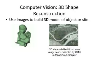
3D reconstruction of a femoral shape using a parametric model and two 2D fluoroscopic images. Ryo Kurazume, Kaori Nakamura, Yumi Iwashita, Tsutomu Hasegawa Graduate School of ISEE, Kyushu University, Japan Toshiyuki Okada, Yoshinobu Sato, Nobuhiko Sugano, Tsuyoshi Koyama

E N D
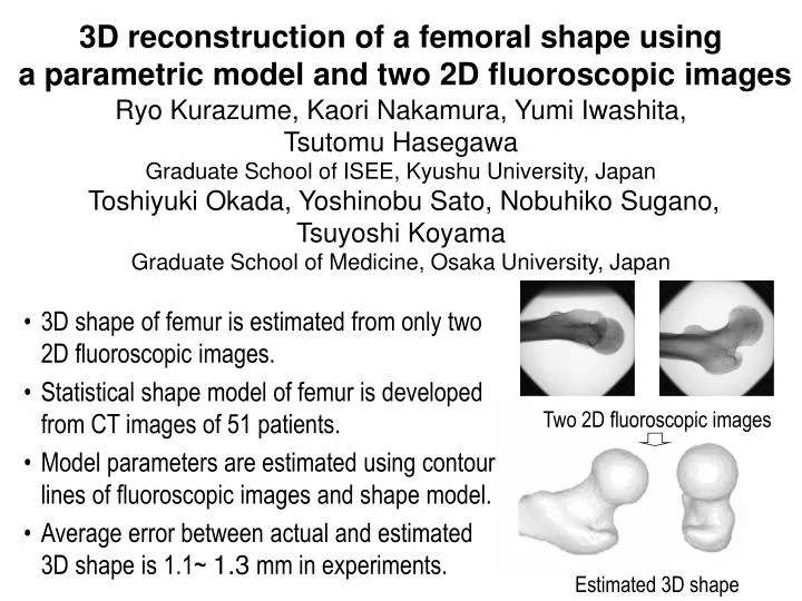
3D reconstruction of a femoral shape using a parametric model and two 2D fluoroscopic images Ryo Kurazume, Kaori Nakamura, Yumi Iwashita, Tsutomu Hasegawa Graduate School of ISEE, Kyushu University, JapanToshiyuki Okada, Yoshinobu Sato, Nobuhiko Sugano, Tsuyoshi Koyama Graduate School ofMedicine, Osaka University, Japan • 3D shape of femur is estimated from only two 2D fluoroscopic images. • Statistical shape model of femur is developed from CT images of 51 patients. • Model parameters are estimated using contour lines of fluoroscopic images and shape model. • Average error between actual and estimated 3D shape is 1.1~ 1.3 mm in experiments. Two 2D fluoroscopic images Estimated 3D shape