Download

1 / 36
370 likes | 607 Vues
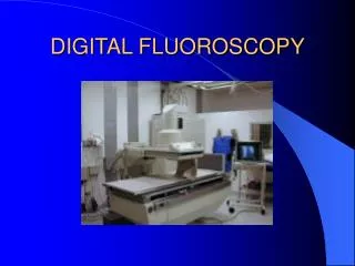
Fluoroscopy State Syllabus. Pages 12, 13; 46, 47; 59-67; 77, 83-96; appendices: 115-120,125. Patient Dose (p. 12). Several factors impact the patient dose Body habitus Type of tissue exposed Tissue density – patient habitus and disease pathology can alter tissue density

E N D
FluoroscopyState Syllabus Pages 12, 13; 46, 47; 59-67; 77, 83-96; appendices: 115-120,125
Patient Dose (p. 12) • Several factors impact the patient dose • Body habitus • Type of tissue exposed • Tissue density – patient habitus and disease pathology can alter tissue density • Elemental composition (atomic number) • The higher the A number of the tissue the more radiation is absorbed • Bone and aluminum have a similar A number (~13) lead is 82
Factors increasing Scatter radiation • High kVp • Large field size (cone wide open) • Thick body part • Large field size is the largest factor
Health Effects of Low-level Radiation Dose (p. 59) • Somatic dose is the dose that the person themselves receives. • This includes an embryo or fetus that is exposed in the womb. • Somatic changes that can occur are: • Injuries to the superficial tissue • Induction of cancer • Cataract formation, impaired fertility, and life-span shortening (the last one really doesn’t exist anymore)
Health Effects of Low-level Radiation Dose (p. 59) • Somatic Dose Indicators are used to measure dose to specific areas of the body. Measurement of one area can’t express the entire somatic dose as: • Primary beam is restricted so measuring a distant point can’t tell you the dose of another area. • Shielding may protect some areas during exposure so measuring the protected area doesn’t give you the dose of the unprotected area • Some natural shielding occurs, some organs cover other tissues.
Health Effects of Low-level Radiation Dose (p. 60) • Somatic Dose Indicators are still useful, however. • Bone Marrow • Bone marrow dose is a reasonable indicator of internal organs that are sensitive to cancer induction (lung, GI tract). • Irradiation of the bone marrow causes hematological depression, particularly to the lymphocytes. • A strong correlation exists between leukemia and the mean radiation dose to the active bone marrow • High bone marrow dose exams are: BE, UGI, and abdominal angio
Health Effects of Low-level Radiation Dose (p. 60) • Thyroid and Skin • The somatic indicator of skin dose of the anterior chest can be a reasonable indicator of breast dose • The skin dose from an esophagram can be indicative of the thyroid dose.
Health Effects of Low-level Radiation Dose (p. 60/61) • Genetic Dose Indicators • The genetic dose is indicated by the damage exhibited by FUTURE offspring of the irradiated person (NOT the embryo or fetus who was themselves irradiated). • When germ cells are irradiated changes may occur in the genes/chromosomes of these cells and be passed on to the descendants of the person who was irradiated
Health Effects of Low-level Radiation Dose (p. 61) • Genetically Significant Dose (GSD) • The GSD is a statistic that helps us estimate the magnitude of genetic effects caused by radiation exposure in a population. • The GSD is defined as the gonad dose which, if received by every member of the population, would be expected to produce the same total genetic effect on the population as the sum of the individual doses that are actually received. (does not include background population • 3 parameters • Number of future children • X-ray exam rate • Mean gonad dose per exam
Biological Effects and Significance of Radiation Dose(p. 62) • When something is irradiated and the energy is transferred to matter ionizations and excitations are produced • Ionizations – electrons removed from an atom • Excitations – electron vacancies in shells • Biological damage from ionizing radiation seems to follow a linear, nonthreshold dose relationship and it is influenced by: • Dose rate to tissue • Total dose received • Type of cell irradiated
Biological Effects and Significance of Radiation Dose (p. 62) • Radiobiological Injury • Cellular amplification – normal cellular metabolic activity can amplify the radiation damage causing the injury to move from the molecular to the microscopic level and resulting in possible gross cellular malfunction • Gross cellular effects – the effect most often seen is the cell stops dividing. It may be temporary or permanent depending on the radiation damage. • Factors seen: Chromosome breaks, clumping of chromatin, abnormal mitoses, increased granularity of cytoplasm, nuclear disintegration, changes in motility or cytoplasmic activity, vacuolization, altered protoplasmic viscosity, changes in membrane permeability
Biological Effects and Significance of Radiation Dose (p. 63) • Radiobiological Injury • Latent Period – where no injury shows up • Early effects – seen in minutes, days, or weeks • Late effects – years, decades, and generations later • Determinants of Biological Effects • 1. Dose-effect curve – a way to graph the dosage administered against the probability of effect. • Threshold –a point where there is no damage seen • Non-threshold – always some effect • Linear – as dose increases effect increases, directly proportional • Non-linear – takes into account fractionation and protraction and other factors • Radiation protection guidelines are based on linear non-threshold
Biological Effects and Significance of Radiation Dose (p. 64) • Determinants of Biological Effects • 2. area exposed and shielding – even partial shielding of radiosensitive blood-forming organs (spleen and bone marrow) can decrease the total radiation effect (especially in children) • 3. Variations in cell sensitivity – radiosensitivity depends on the • number of undifferentiated cells in the tissue • degree of mitotic activity in tissue • length of time cells stay in active prolifereation
Biological Effects and Significance of Radiation Dose (p. 64/65) • Determinants of Biological Effects • 4. Short-term effects • in general at 25 rads or less there are no indications of injury (based on animal experimentation for the most part) • 5. Long-term effects – manifest years later. • Can be from acute or chronic low-level exposure. • Long-term effects of low level dose on many people is more of a health concern than high dose on a few people • No unique disease associated with long-term effects they are measured by statistic increase in some diseases • Observed: somatic changes such as cancer, embryological effects, cataracts. Genetic mutations that show up generations later
Biological Effects and Significance of Radiation Dose (p. 65) • Determinants of Biological Effects • 6. carcinogenic effects – human evidence that radiation contributes to induction of cancer • Early radiologists and dentists manifested an increase in skin malignancies and leukemia • Radium dial painters showed increase in bone malignancies • Uranium miners increase in lung cancer • Japanese survivors of Hiroshima/Nagasaki increase in leukemia and cancer • Frequent radiation-induced cancers: • Female breast, thyroid gland (women/children most), hemopoietic tissue, lungs, GI tract, bones
Biological Effects and Significance of Radiation Dose (p. 66) • Determinants of Biological Effects • 7. Embryological effects • highly sensitive to radiation (immature, undifferentiated, rapidly dividing cells) • 50 rad to fetus can result in spontaneous abortion • 8. Cataractogenic effects • Several hundred rads of acute dose • Can tell if the cataract was caused by radiation • 9. Life-span shortening • Really doesn’t exist in today’s world
Biological Effects and Significance of Radiation Dose (p. 66/67) • Determinants of Biological Effects • 10. Genetic Effects • Gonads/germ cells can be damaged by radiation and the damage is then passed on to future offspring and can alter their genetic code • Most genetic mutations are harmful, the more harmful they are the quicker they will disappear. • Animal experimentation tells us that: • No indication of a threshold dose for genetic effects of radiation • Mutations appear to be dose-rate dependent. If the dose-rate is lower and protracted less damage is done
Visual Physiology (p. 77) • Cones • Daylight vision • Photopic vision • Perceive color • Concentrated in center of retina • Visual acuity good • Rods • Nighttime vision • Scotopic vision • Perceive grays • Concentrated on the periphery of the retina • Visual acuity poor View “eye” on page 77 Be able to label eye
Visual Physiology (p. 77) • Visual acuity • The ability to perceive fine detail • Integration time • The time required by the eye for recognition of an image is 0.2 second • Normal viewing distance • 12-15 inches • The retina receives the images of external objects • The blind spot • Where the optic nerve enters the retina and there are no rods or cones • The Macula Lutea is the exact center of the retina • There is a great increase in the number of cones and it is the region of greatest visual acuity
Pediatric Fluoroscopy (p. 46) • Doses given to children are much lower than doses that adults receive, however, it can be more dangerous as: • their lifespan is longer so there is more time for more radiation dose to occur and for damage to manifest itself • Children are more sensitive to radiation as their tissues are young and rapidly dividing as they grow (increased mitotic activity)
Pediatric Fluoroscopy (p. 46) • The reason for repeats on children stems from motion the majority of the time. You can decrease motion by: • Being friendly and nonthreatening • Having the parents hold the child • Secure children by mechanical means • Protection for children • Gonadal shielding is a must whenever the exam will not be compromised. • Avoid contrast-filled structures centering over phototimer/Automatic Brightness Control as this will increase patient dose
Appendix 5 (p. 83) • Review appendix 5 for your own benefit and understanding.
Appendix 6 (p. 84) • The Pregnant Patient • The interruption of pregnancy is never justified because of radiation does to embryo/fetus from a diagnostic x-ray exam, including abdominal exams • Postpone exams of pregnant patients whenever possible
Appendix 7 (p. 85/86) • Occupationally Exposed Women • A declared pregnant woman is one who has voluntarily informed her employer, in writing, that she is pregnant • The dose equivalent to an embryo/fetus (during the entire pregnancy) due to occupational exposure should not exceed 0.5 rem (5 mSv) • The radiation dose of the embryo/fetus shall not be greater than 0.05 rem in any month (occupational exposure)
Appendix 12 (p. 115-117) Review images in syllabus • Isoexposure curves • The main source of scattered radiation during fluoroscopy is the patient • Operator exposure to scattered radiation is directly proportional to patient exposure and is influenced by: • kVp, area exposed, area exposed, thickness of body part, time of exposure • When the X-ray tube is located under the table there is less exposure to the patient. • Most of the exposure is under the table at angles of 135 and 120 degrees from the primary beam
Appendix 13, 14, 15 • Review Appendices 13,14, 15 on pages 118, 119 and 120. • These appendices tell you: • approximate organ doses by examination • somatic detriment from common x-ray exams • possibilities for dose reduction
Appendix 17 (p. 122) excerpts • All hospitals shall maintain diagnostic radiological services • All persons operating or supervising the operation of X-ray machines shall comply with the requirements of the Regulations Relating to Radiologic Technology • Diagnostic radiological services may be performed on the order of a person lawfully authorized to give such an order • X-ray films or reproductions thereof, shall be retained for the same period of time as is required for other parts of the patient’s medical record
Appendix 17 (p. 122) excerpts • A physician shall have overall responsibility for the radiological service. • Sufficient certified radiologic technologists shall be employed to meet the needs of the service being offered • There shall be at least one person on duty or on call at all times capable of operating radiological equipment • There shall be sufficient equipment and supplies and space to adequately provide radiological services.
Appendix 18 (p. 124) • Review appendix 18 on p. 124 to see the primary fluoroscopic beam attenuation factors.
Appendix 19 (p. 125) • Personnel Monitoring Devices • Film Badge • Detects x-rays, gamma, beta, thermal neutrons, fast neutrons • Range: 0.0-700 rad • Detects 10keV for gamma and 200 keV for beta • Advantages: inexpensive, estimates of integrated dose, permanent records, objective review, detects problems • Disadvantages: moderate directional dependence, strong energy dependence for low energy x-rays, false readings produced by heat, pressure, vapors
Appendix 19 (p. 125) • Personnel Monitoring Devices • Thermoluminescent Dosimeter (TLD) • Detects x-rays, gamma, beta, thermal, neutrons, fast neutrons • Range: 10 mrads to 10 5rad • Detects 10 keV minimum • Advantages: infinite shelf life, small size, low directional dependence, reusable, inexpensive, integrated dose estimates • Disadvantages: cancellation of dose upon reading, dose range depends on sensitivity of reader, increased sensitivity with each use, subjective info
Appendix 19 (p. 125) • Personnel Monitoring Devices • Pocket Ionization Chamber • Detects: x-rays, gamma, beta, thermal neutrons, fast neutrons • Range: 0.001 to 2000 rads (theoretical), x-rays 0.001-200 millirads • Detects: 10 keV for gamma, 20keV for fast neutrons minimum • Advantages: accurate info quickly, small size, low directional dependence, economical, little maintenance, reusable • Disadvantages: no permanent record, frequent reading tabulation, recharging may be required, subject to accidental discharge (shock, electrical leakage, range of measurement is limited
Appendix 12 - Isoexposure Curves • Protective aprons do not eliminate all exposure • 0.25 mm lead equivalent eliminates 96% while 0.5 mm eliminates 99%
Miscellaneous Facts • Remnant radiation, that emerges from the patient, and forms them image, is only 5% of the incident photons • The minimum source-to-skin distance for portable fluoroscopic equipment is 12” and should be at least 18” for stationary fluoroscopic equipment • P. 60 50 rads, in one dose, can result in cessation of sperm formation, however, fertility is not impaired until preexisting sperm are eliminated which takes weeks. It is a temporary impairment not permanent sterility
Miscellaneous Facts • Females have all of their germ cells, or ooctyes and can not replace them as the male replaces sperm. • Ooctyes are radiosensitive as irradiation causes a lasting reduction in reproductive potential. 30 rads in one dose can cause temporary sterility. • These doses, to the gonads, should not be encountered in the diagnostic radiology arena.