Download
1 / 8
190 likes | 611 Vues
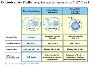
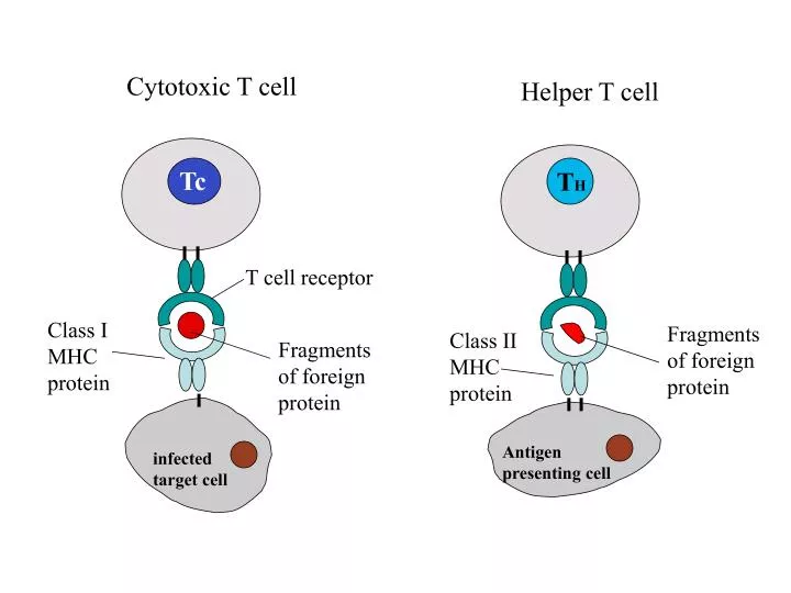
Cytotoxic T cell. Helper T cell. T H. Tc. T cell receptor. Fragments of foreign protein. Class II MHC protein. Class I MHC protein. Fragments of foreign protein. Antigen presenting cell. infected target cell. Helper T cells: • Express CD4 molecule on the cell surface.

E N D
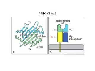
Cytotoxic T cell Helper T cell TH Tc T cell receptor Fragments of foreign protein Class II MHC protein Class I MHC protein Fragments of foreign protein Antigen presenting cell infected target cell
Helper T cells: • Express CD4 molecule on the cell surface. • Recognize antigen peptides bound to class II MHC molecules (exogenous or ingested antigens). • Provide help to B cells, macrophages and other T cell effector responses though the production of cytokines Cytotoxic T cells: • Express CD8 molecule on the cell surface. • Recognize antigen peptides bound to class I MHC molecules (endogenously produced antigen such as viral antigen). • Kill infected target cells.
IL-2 Gene +1 +47 -286 -257 -256 -242 -208 -188 -158 -145 -93 -66 TATA NF-AT NFIL-2D NFIL-2C NFIL-2B NFIL-2A NFAT Z Construct -70 +1 +47 TATA lac Z TK promoter Hygromycin Resistance gene NF-AT NF-AT NF-AT IL-2
Detection of ß-Galactosidase: Upon addition of its chromogenic substrate, chlorophenol red galactoside (CPRG), ß-galactosidase hydrolyzes CPRG, turning it from a yellow color to purple at alkaline pH. The color changs thus provide a convenient measure of gene transcription activities during T cell activation.
2+ Ca Receptor CRAC 2+ Ca IP3R Ras, Rac, PKC PO3 CsA/Cph NF-ATc Calcineurin NF-AT kinase NF-ATn NF-ATc IL-2 T cell receptor activation Ca2+ NF-ATc IP3 Phosphoinositol pathway IL-2 DAG PKC NF-ATn
2+ Ca 2+ Ca PO3 NF-ATc Con A EGTA Receptor CRAC Ionomycin thapsigargin IP3R Ras, Rac, PKC CsA/Cph PMA Calcineurin NF-AT kinase NF-ATn NF-ATc IL-2
1. Mammalian cell culture: Goal: learn how to count cells using a hemacytometer Learn to grow mammalian cell lines 2. Signal transduction lab: Examine the effects of various inhibitors and activators on IL-2 expression. This week’s experiments
How to count cells using a hemacytometer? Cell unit per box: 1 x 10⁴ cells/ml Blue cells : 3 x 10⁴/ml