Download
1 / 50
540 likes | 665 Vues
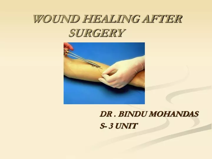
WOUND HEALING AFTER SURGERY. DR . BINDU MOHANDAS S- 3 UNIT. Definition:. Wound healing is a mechanism where by the body attempts to restore the integrity of the injured part. The process of healing involves two distinct process 1 . REPAIR 2 . REGENERATION. REPAIR.

E N D
WOUND HEALING AFTER SURGERY DR . BINDU MOHANDAS S- 3 UNIT
Definition: Wound healing is a mechanism where by the body attempts to restore the integrity of the injured part.
The process of healing involves two distinct process 1 . REPAIR 2 . REGENERATION
REPAIR • When healing takes place by proliferation of connective tissue elements. • It results in fibrosis and scarring
REGENERATION • When healing takes place by proliferation of parenchyml cells • It usually results in complete restoration of original tissues • Tissues with high proliferative capacity can regenerate after injury as long as the stem cells of these tissues are not destroyed
Takes place in 3 phases 1. Inflammatory phase 2. Proliferative phase 3. Remodelling phase -
Inflammatory phase • Begins immediately after wounding • Lasts for 2-3 days. • PMN cells & macrophages are attracted towards the fibrin clot.
Proliferative phase • lasts from 3rd day to 3rd week • Consist mainly of fibroblast activity with production of collagen & ground substance, angiogenesis & reepithelisation of wound surface.
Remodelling phase • Maturation of collagen • Wound strengthening occurs
Skin wounds are classically described to heal by A) by primary intention B) by secondary intention • This is based on the nature of the wound rather than the healing process.
Healing by first intention • Wound with opposed edges. • Healing of a clean, uninfected surgical incision approximated by surgical sutures. • Also called as healing by primary union.
Steps:- • Within 24 hrs – neutrophils appears at the margins of the incision, moving towards the fibrin clot. • 24-48 hrs – spurs of epithelial cells move from the wound edges along the cut margins of the dermis. Depositing the basement membrane components.
By 3rd day – neutrophils are replaced by macrophages – granulation tissue invades the incision and collagen fibres appears near the margins of the incision – granulation issue invades the incision and collagen fibres appears near the margins of the incision – epithelial cell proliferation thickens the epidermal layer.
By day 5- incisional space is filled with granulation tissue, with maximal neovascularization- collagen fibrils are abundant & bridges the incision.- epidermis recovers its normal thickness
By 2nd week – continued accumulation of collagen and proliferation of fibroblasts within the incisional scar, accompained by regression of vascular channels. • By end of 1st month- the scar is made up of cellular connective tissue devoid of inflammatory infiltrate, the decimal appendages are permanently lost in the line of incision. Tensile strength of the wound increases.
Healing by secondary intention (Secondary union) Occurs in wounds with separated edges. Inflammatory reaction is more intense. Large amount of granulation tissue are formed wound contraction-reduces the size of the wound Healing takes place from the base to upwards as well as from the margin inwards
Delayed primary intention (teritiary intention) • Wound is initially left open • Edges are later opposed when healing conditions are favourable
1) Bone • Periosteal and endosteal proliferation leads to callous formation • In remodelling phase, cortical structure & the medullary cavity are restored.
2) Nerve • Distal to the wound, Wallerian degeneration occurs • Proximally the nerve suffers traumatic degeneration as far as the last node of Ranvier • Nerve regeneration is characterised by profuse growth of new nerve fibres which sprout from the cut proximal end.
3)Muscles • SKELETAL MUSCLE - similar to peripheral nerve regeneration - on injury, cut ends of muscle fibres retract - the injured site is filled with fibrinous material, polymorphs & macrophages
SMOOTH MUSCLE - It has limited regenerative capacity - In large destructive lesions, smooth muscle is replaced by permenant scar tissue • CARDIAC MUSCLE -Destruction of heart muscle is replaced by fibrous tissue
HEALING OF MUCOSAL SURFACE • Very good regenerative power - Occurs by proliferation from margins, migration, multilayering, and differentiation of epithelial cells
HEALING OF SOLID EPITHELIAL ORGANS - Following gross tissue damage to organs like kidney, liver & thyroid, the replacement is by fibrous scar - But if the basement membrane is intact & only parenchymal damage is present, regeneration occurs
Follow principles of ALTS • Thorough examination of wound site after copious saline irrigation • Examine the possible structures that are damaged under analgesia • Assess the movement and sensation • wound exploration and diagnosis
A bleeding wound should be elevated & pressure pad is applied • WOUND DEBRIDEMENT- wound should be debrided to limit of blood supply, devitalised tissues must be excised • Repair of all damaged structures are attempted
1. Bites • As per above • Antibiotic coverage for aerobic and anaerobic orginisms
2. Haematomata • Release by incision/aspiration • Surgical exploration is case of calcification
3. Degloving • Open – ring avulsion injury with loss of finger skin • Closed – rollover injury
4. Compartment Syndrome • Occurs in closed lower limb injuries • Characterised by severe pain, pain on passive movement of the affected compartment muscles, distal sensory disturbance, absence of pulse distally. • treatment: fasciotomy
5. High pressure injection injuries • Occurs in person working with cleaning, degreasing and painting devices • tissue damage depends on toxicity of substance and injection pressure • Treatment is surgical with wide exposure, removal of the toxic substance and through debridement.
1. Leg ulcers • Treatment of the underlying cause • A chronic ulcer unresponsive to treatment should be biopsied to rule out neoplastic change • Surgical treatment is required if non operative treatment fails • Meshed skin grafts may be required
2. Pressure sores • Prevention – good skin care foam beds pt. turning at least every 2hrs • Preoperative treatment of pressure sore involves adequate debridement and the use of vaccum assisted closure.
1. Local factors Blood supply Mechanical stress denervation Necrotic issue Local infection Protection (dressing) Foreign Body Surgical techniques Hacmatoma type of tissue
2. Systemic factors Age Malnutruction Anemia obesity Drugs (steroids, cytotokie, medications) Systemic infection genetic disorders temperature Hormones trauma, Hypevolemia hypoxia Diabetes Vit & trace metal deficiency Malignant disease.
Hypertrophic scar & keloid • Wound dehisense & ulceration • Contractures • Wound infection • Incisional hernia • Implantation cyst formation • Pigmentation • Neoplasia
Source of information • Robin’s text book of pathology • Harsh mohan’s text book of pathology • Bailey and love’s book of surgery • Sabiston’s book of surgery • www.pubmed.com • www.google.com