Download
1 / 39
430 likes | 600 Vues
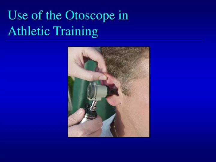
Use of the Otoscope in Athletic Training. Objectives. Briefly discuss the types and features of the otoscope Provide an overview of otoscopic assessment procedures Present a clinical teaching model for teaching your students to properly use the otoscope

E N D
Objectives • Briefly discuss the types and features of the otoscope • Provide an overview of otoscopic assessment procedures • Present a clinical teaching model for teaching your students to properly use the otoscope • Provide educational resources for teaching otoscopy
Types of Otoscopes • Pocket style • < $50 • Clinical model • $200 - $400+ Pocket style Clinical model
Features of the Otoscope • Power source • Battery (most common in athletic training clinical setting) • Electric • Light source • Incandescent bulb (produces a yellow light) • Hallogen bulb (best – produces a white light)
Features of the Otoscope • Magnifier • Not available on all models • Provides better view of tympanic membrane, particularly for beginners
Features of the Otoscope • Speculum • Variety of sizes • Reusable or disposable
Examination of the Ear • History • Observation • Palpation • Specialtests • Otoscopic assessment
Examination of the Ear • History • Trauma • Allergies, colds, sinus drainage • Changes in pressure (flying, diving) • Dizziness • Changes in hearing • Duration of symptoms
Examination of the Ear • Observation • Redness • Swelling • Drainage • Foreign object • Cuts, scrapes, bruises
Examination of the Ear • Palpation • Gentle pressure on tragus
Examination of the Ear • Palpation • Traction on ear lobe & pinna
Otoscopic Assessment • Evaluate the noninvolved ear first • This practice provides a basis for comparison AND prevents cross-contamination
Otoscopic Assessment • Step 1: • Place your patient in a seated position with his/her head turned slightly downward and away from the ear to be examined
Otoscopic Assessment • Step 1 (cont.): • I teach this as the “puppy position” (puppies always cock their heads to the side when you talk to them)
Otoscopic Assessment • Step 2: • Select the largest possible speculum that can be comfortably inserted into the ear
Otoscopic Assessment • Step 2 (cont.): • When inserted, the speculum should fit snugly in the outer third of the canal and rest against the tragus and anterior wall of the canal Modified from Middle Ear Conditions. Anatomical Chart Co. Skokie, IL, 1999.
Otoscopic Assessment • Step 2 (cont.): • Choosing a speculum that is too small will cause movement within the canal • Excessive movement can cause discomfort for your patient Modified from Middle Ear Conditions. Anatomical Chart Co. Skokie, IL, 1999.
Otoscopic Assessment • Step 3: • Hold the otoscope with the same hand as the ear you are examining • right ear, right hand • left ear, left hand
Otoscopic Assessment • Step 3 (cont.): • The otoscope should be stabilized by placing the ring and little finger resting on the patient’s cheek or temple
Otoscopic Assessment Pencil Grip Hammer Grip
Otoscopic Assessment • Step 4: • Pull the pinna upward and backward to straighten the canal Modified from Middle Ear Conditions. Anatomical Chart Co. Skokie, IL, 1999.
Otoscopic Assessment • Step 5: • While maintaining traction on the pinna, place the speculum of the otoscope at, but not in the ear canal
Otoscopic Assessment • Caution: • Never insert the otoscope blindly • Always“Watch your way in”
Otoscopic Assessment • Tip: • If the patient experiences pain, reposition the canal by adjusting the angle and degree of traction on the pinna
Otoscopic Assessment • Caution: • If the patient’s discomfort persists even after readjustment of the canal, halt the examination and refer the patient to a physician.
Otoscopic Assessment • Step 6: • Once the tympanic membrane comes into view, rotate the speculum to view as much of the membrane as possible • Posterior superior • Anterior superior • Anterior inferior • Posterior inferior Marty DR. The Ear Book. Jefferson City, MO: Lang ENT Publishing. 1987;Color plate 1.
Otoscopic Assessment • Tip • Like trying to view the corners of a room through a key hole Marty DR. The Ear Book. Jefferson City, MO: Lang ENT Publishing. 1987;Color plate 1. Modified from Middle Ear Conditions. Anatomical Chart Co. Skokie, IL, 1999.
Otoscopic Assessment • Tip Fincher AL. Use of the otoscope in the evaluation of common injuries and illnesses of the ear. J Athl Train. 1994;29:53,54. • The posterior inferior portion of the membrane is often difficult to see This is due to the angle of the membrane within the canal Modified from Middle Ear Conditions. Anatomical Chart Co. Skokie, IL, 1999.
Otoscopic Assessment • Step 7: • Inspect the membrane for color, clarity, & position • Pearly gray • Semitransparent • Not bulging or retracted L R Fincher AL. Use of the otoscope in the evaluation of common injuries and illnesses of the ear. J Athl Train. 1994;29:53,54.
Short process Umbo Otoscopic Assessment • Step 8: • Identify key landmarks • Malleus • Manubrium • Short process • Umbo L R • Light reflex Fincher AL. Use of the otoscope in the evaluation of common injuries and illnesses of the ear. J Athl Train. 1994;29:53,54.
Otoscopic Assessment • Step 8 (cont.): • Identify key landmarks • Note that manubrium angles toward the 10:00 position in the left ear and the 2:00 position in the right ear L R Fincher AL. Use of the otoscope in the evaluation of common injuries and illnesses of the ear. J Athl Train. 1994;29:53,54.
Pars flaccida Pars tensa Otoscopic Assessment • Step 8 (cont.): • Identify key landmarks • Pars flaccida • Pars tensa L R • Annulus Fincher AL. Use of the otoscope in the evaluation of common injuries and illnesses of the ear. J Athl Train. 1994;29:53,54.
Otoscopic Assessment • Step 8 (cont.): • Identify key landmarks • Look beyond the membrane • Stapes • Incus Fincher AL. Use of the otoscope in the evaluation of common injuries and illnesses of the ear. J Athl Train. 1994;29:53.
Otoscopic Assessment • Step 9: • Look for abnormalities • Fluid • Perforations Fluid & Air Bubbles Fincher AL. Use of the otoscope in the evaluation of common injuries andillnesses of the ear. J Athl Train. 1994;29:54. Perforation Marty DR. The Ear Book. Jefferson City, MO: Lang ENT Publishing. 1987;Color plate 8.
Otoscopic Assessment • Step 10 • Work with your team physician to develop your confidence and skill • PRACTICE, PRACTICE, PRACTICE !!! • You must look at many ears to develop to become comfortable with “normal”
Guided, Self-Directed Activities – Post Lab • Content • Recognition of pathology – visual images Perforation Middle ear fluid Marty DR. The Ear Book. Jefferson City, MO: Lang ENT Publishing. 1987;Color plate 8. Marty DR. The Ear Book. Jefferson City, MO: Lang ENT Publishing. 1987;Color plate 3.
Guided, Self-Directed Activities – Post Lab • Content • Recognition of pathology – visual images Perforation Otitis Media Modified from Middle Ear Conditions. Anatomical Chart Co., Skokie, IL. 1999. Modified from Middle Ear Conditions. Anatomical Chart Co., Skokie, IL. 1999.