Download
1 / 59
630 likes | 1.47k Vues
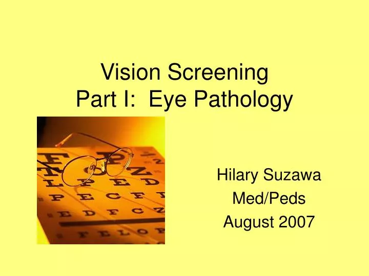
Vision Screening Part I: Eye Pathology. Hilary Suzawa Med/Peds August 2007. Frequency. Vision problems in 5-10% of preschoolers 3 main problems Refractive Error Strabismus 4% preschoolers Amblyopia Strabismus is most common cause of amblyopia

E N D
Vision ScreeningPart I: Eye Pathology Hilary Suzawa Med/Peds August 2007
Frequency • Vision problems in 5-10% of preschoolers • 3 main problems • Refractive Error • Strabismus • 4% preschoolers • Amblyopia • Strabismus is most common cause of amblyopia • 40% of those with Strabismus develop amblyopia • Leading cause of monocular vision loss in 20-70 yo
Refractive Error • Focusing problems • Myopia (nearsightedness) • Eyeball too long • Image focuses in front of retina • Hyperopia (farsightedness) • Eyeball too short • Image focuses behind the retina • Astigmatism • Cornea is misshapen
Myopia Eyeball too long Image focuses in front of retina Correct with concave lens Hypermetropia Eyeball too short Image focuses behind retina Correct with convex lens Astigmatism Cornea is misshapen Correct with cylindrical lens
Strabismus • Misalignment of the eyes • Timing • All the time (tropia) • Temporary (phoria) • Direction • Eso (inwards; nasally) • Exo (outwards; temporally) • Hyper (upwards; towards the sky) • Hypo (downwards; towards the ground)
Strabismus • Medial (eso) deviations are the most common ocular deviations • Account for >50% of all cases of strabismus
Pseudostrabismus • Appearance of misalignment of the eyes • Broad nasal bridge covers the nasal sclera of one or both eyes • Check for symmetry of light reflection
Infantile Strabismus • Severe esotropia before age 6 months • +FMH strabismus • Refraction of each eye often normal • Surgical realignment before age 2 years
Accommodative Esotropia • Most children normally have mild hyperopia (farsighted) • Severe hyperopia may lead to accommodative esotropia
Accommodative Esotropia • In order to overcome the refractive error (hypermetropia) the eye accommodates leading to medial deviation • Often hereditary • Average onset at age 2-3 years • Glasses to correct
Nonaccommodative Esotropia • Results from ocular insults • Trauma • Prematurity • Cataract • Neurodevelopmental abnormalities • Syndromes affecting CN VI (lateral rectus) • Mobius Syndrome • Duane Syndrome
Mobius Syndrome Congenital Facial Diplegia Under-development of CN VI and CN VII
Duane Syndrome Under-development of nucleus for CN VI Inability for lateral gaze Retraction of the eye
Exo Deviations • 25% of ocular misalignments • Often hereditary • Manifest intermittently with fatigue, illness, distance fixation (exophoria) • Suspect ocular or CNS pathology if late-onset exotropia
Vertical Deviations • <10% ocular misalignments • Often compensatory head tilt to minimize diplopia • CN IV paresis • Brown Syndrome—defect in superior oblique muscle • Retro-orbital tumors • Thyroid ophthalmopathy • Facial Trauma
Brown Syndrome Superior Oblique Tendon Sheath Syndrome Restriction of eye elevation with adduction
Amblyopia • Loss of visual acuity due to active cortical suppression of the vision of an eye
Causes of Amblyopia • Strabismus—misalignment • Anisometropia—one eye has a different length from the other resulting in a different focusing ability (refractive error) • Visual deprivation—not using an eye
Causes of Amblyopia • Strabismus • Esotropia, Exotropia, Hypertropia • Anisometropia • Hyperopia, Astigmatism, Aniseikonia (unequal retinal images—rare) • Visual Deprivation • Unilateral: Cataract, ptosis, opaque cornea, hyphema, prolonged patching, prolonged use atropine drops • Bilateral: Cataract, nystagmus
Hyphema Blood in anterior chamber of the lens
Treatment • Surgical realignment of strabismus • Patching of the good eye • Cycloplegic drops to decrease visual acuity of the good eye
Bibliography • Broderick, Peter. Pediatric Vision Screening for the Family Physician. American Family Physician 1998; 58 (3). • Tingley, Donald. Vision Screening Essentials: Screening Today for Eye Disorders in the Pediatric Patient. Pediatrics in Review. 2007; 28 (2): 54-61.
Vision ScreeningPart II: Clinical Evaluation Hilary Suzawa Med/Peds August 2007
Clinical Screening • Goal of screening is to preserve vision • Birth-3 years • Check anatomy and gross visual assessments • >3 years • Start visual acuity screening
Infants • External penlight exam for abnormalities of eye and surrounding tissues • Ocular alignment (corneal reflections) • Red Reflex
Infants • Red Reflex • Equal in brightness and color
Abnormal Red Reflex • Abnormal (Leukocoria) • Anisometropia (varying eye length) • Refractive error • Cataract • Retinoblastoma • If abnormal Red Reflex, refer to ophtho
Infants • Funduscopic exam to evaluate Retinopathy of Prematurity (ROP)
Infants • Check if equal responsiveness to light stimulus • Visual acuity 20/400 at 1 month and improves with age to 20/20 by age 8 years
6 months • Ability to fix and follow light, face, or small toy • External penlight exam for abnormalities of eye and surrounding tissues • Pupil exam • Ocular alignment (corneal reflections) • Red Reflex
3-4 years • Visual acuity by appropriate eye chart (picture or tumbling E) • External penlight exam for abnormalities of eye and surrounding tissues • Pupil exam • Ocular alignment (ocular movements, cover test and corneal reflections) • Red Reflex and +/-fundus exam
5-6 years • Visual acuity by appropriate eye chart (Snellen) • External penlight exam for abnormalities of eye and surrounding tissues • Pupil exam • Ocular alignment (ocular movements, cover test and corneal reflections) • Red Reflex and +/-fundus exam
Visual Acuity • Use age-appropriate chart • 10 ft from the chart • One eye tested at a time • Make sure pt not able to cheat
Visual Acuity • All children >8 years old should be able to achieve 20/20 VA using their best eyeglass correction • Abnormal • Differences of two lines of visual acuity between the eyes • Less than 20/40 in either eye
Strabismus Testing • Tropia—full-time eye misdirection • Phoria—tendency for the eye to turn when disturbances in binocularity occur (eg one eye is covered) • Distinguish using Cover/Uncover Testing
Cover Testing • Patient fixates on a target • Place a cover over one eye • Normal—uncovered eye does not move • Abnormal—uncovered eye moves to look at target tropia • If the eye moved nasally (exotropia) • If the eye moved temporally (esotropia)