Download
1 / 41
420 likes | 699 Vues
Early Pregnancy Problems. Feras Izzat Consultant Gynaecologist – EGU/EPAU Lead University Hospitals Coventry & Warwickshire NHS Trust. Introduction. Ectopic Pregnancy Bleeding in early pregnancy and miscarriage Gestational Trophoblastic Disease. Ectopic Pregnancy. Definition.

E N D
Early Pregnancy Problems Feras Izzat Consultant Gynaecologist – EGU/EPAU Lead University Hospitals Coventry & Warwickshire NHS Trust
Introduction Ectopic Pregnancy Bleeding in early pregnancy and miscarriage Gestational Trophoblastic Disease
Definition Pregnancy occurring outside uterine cavity Approx 11/1000 of pregnancies – rate increasing Maternal mortality in 1/2500 ectopic pregnancies (11 deaths in most recent report)
Site Tubal Interstitial 2.4% Isthmic 12% Ampullary 70% Fimbrial 11.1% Non Tubal Ovary Abdominal cavity Cervix CS Scar
Risk factors Previous PID Previous ectopic pregnancy Previous tubal surgery (e.g. sterilisation, reversal) Pregnancy in the presence of IUCD POP ART (IVF)
Symptoms Acute Low abdominal pain – peritoneal irritation by blood Vaginal bleeding – shedding of decidua Shoulder tip pain – referred from diaphragm Fainting - hypovolaemia Chronic (Atypical) Asymptomatic, gastrointestinal symptoms
Signs Abdominal tenderness Adnexal tenderness / mass Shock – tachycardia, hypotension, pallor None
Diagnosis Ultrasound Empty uterus, adnexal mass, free fluid, occasionally live pregnancy outside of uterus Serum βhCG & Progesterone Laparoscopy
Ultarsound Trans-Vaginal Ultrasonography Sensitivity 100%, specificity 98.2%. The positive predictive value 98%, and the negative predictive value was 100% FH seen in 23% Timor-Tritschet al, 1990 Am J Obstet Gynecol.
Management Conservative hCG <1000 , Progesterone < 5 stable, success 70% Medical Methotrexate –hCG <4000 mass < 3cm, success 84%. Susequent IUP 54% recurrent EP 8% Surgical - Laparoscopy Salpingectomy, IUP 38.3%, EP 9.8 Salpingotomy, IUP 61.1%, EP 15.5 Yao et al, Fertility Sterility 1997
PUL Pregnancy of unknown location (PUL) - positive pregnancy test with no signs of intra- or extrauterine pregnancy on transvaginal sonography (TVS). 15-20% of all EPAU scans Management should be expectant if stable with an initial serum progesterone (<20) and a hCG ratio 0h/48h of <0.87 Condous et al, Ultrasound Obstet Gynecol 2006
Definitions Threatened miscarriage Vaginal bleeding at < 24 weeks gestation Delayed (silent) miscarriage Gestational sac with/without fetus present (but no FH) Recurrent miscarriage 3 or more consecutive miscarriages (with or without a known cause)
Miscarriage Approximately 30% of pregnant women will experience bleeding in early pregnancy At least 50% of women with threatened miscarriage will have continuing pregnancy Miscarriage occurs in 15-20% of clinically diagnosed pregnancies
Causes of miscarriage Genetic abnormalities 85% Maternal illness e.g. diabetes, Thyroid disease Phospholipid / Lupus – 15% recurrent miscarriages Uterine abnormalities ‘Cervical incompetence’ Progesterone deficiency?
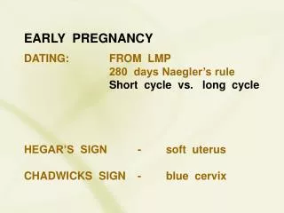
History • LMP • When? • Amount? • Pain? • Timing of Pain
Examination • ABC (vital signs) • Abdominal • Vaginal (speculum) • Cx state • Amount of bleeding
Investigations Ideally in dedicated ‘Early Pregnancy Assessment Unit’ Ultrasound Measurement of serum βhCG Determination of blood & Rhesus group FBC, G&S and admit if significant bleeding Psychological support
Ultrasound Expect to see viable fetus from around 6.5 weeks transabdominally, 5.5 weeks transvaginally Diagnosis can be made on TVS only CRL ≥ 7mm Empty GS with a mean diameter ≥ 25 mm
Measurement of βhCG Not necessary if diagnosis unequivocal on scan Useful as part of investigations to diagnose / exclude extrauterine pregnancy Doubling time approx 2 days in viable pregnancy Halving time 1-2 days in complete miscarriage Should see fetal pole with βhCG of 1500-2000
Management of incomplete miscarriage Conservative 76% success Medical mifipristone & misoprostol – 82% success Nielsen et al, BJOG 1999 Surgical (ERPC) No difference in satisfaction rate than medical – 95% Chipchase et al, BJOG 1995
Recurrent miscarriage Loss of 3 or more consecutive pregnancies Affects 1% of women in reproductive age group Investigations can identify up to 50% with a cause Women aged <=30 years have a subsequent miscarriage rate of 25% which rises to 52% in women aged >=40 years. The risk of a subsequent miscarriage is 29% after 3 miscarriages, this rises to 53% in 6 or more previous miscarriages Clifford et al, Human Reproduction 1997
GTD • The abnormal proliferation of gestational trophoblast tissue • Spectrum of disease • Pre-Malignant • Partial Molar Pregnancy • Complete Molar Pregnancy • Malignant • Invasive mole • Choriocarcinoma • Placental site trophoblastic tumours
Molar Pregnancy 1 in 1000 live births Partial Partial moles are triploid with 2 sets of paternal and 1 set of maternal chromosomes An embryo often present that dies at 8-9 weeks 0.5% need chemotherapy for invasive disease Complete No fetal pole, diplod chromosomes paternally derived – androgenetic No embryo Chemo therapy rate 8-20%
Presentation Vaginal bleeding Excessive N&V ‘Hyperemesis gravidarum’ Uterus large for dates
Diagnosis Ultrasound Histology after surgical evacuation
Follow-up Monitor via regional centre – London, Sheffield, Dundee CM – 8-20% risk of invasive disease PM – 0.5% Choriocarcinoma may follow any subsequent pregnancy – miscarriage, TOP, term delivery Choriocarcinoma is curable Monitor βhCG levels to check resolution – for 6 months to 2 years