Download

1 / 28
370 likes | 629 Vues
Development of The Eye And Ear. Dr. Nimir Dr. Safaa. Objectives Describe the development of the optic cup, and lens, Describe the development of the retina, ciliary body & iris. Describe the development of choroid ,sclera and cornea.

E N D
Development of The Eye And Ear Dr. Nimir Dr. Safaa
Objectives • Describe the development of the optic cup, and lens, • Describe the development of the retina, ciliary body & iris. • Describe the development of choroid ,sclera and cornea. • Describe the development of the optic nerve and vitrousbody. • List the anomalies associated with eye development • Discuss the clinical bases of the above anomalies • Describe the development of inner ear • Describe the development of middle ear • Describe the development of the external ear • List the anomalies might be associated with ear development • Discuss the clinical cases of the ear anomalies
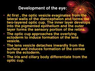
Development of Eye • Optic Cup and Lens Vesicle: • Developing eyes appear in the 22-day embryo as an optic grooves on sides of forebrain. With closure of neural tube, these grooves form optic vesicles. • Optic vesicle invaginates and forms optic cup. This invagination inferiorly forms choroid fissure which allows hyaloid artery to reach inner chamber of the eye. • During seventh week, lips of choroid fissure fuse & becomes future pupil.
Optic vesicles come in contact with ectoderm and induce formation of lens placodewhich invaginates and develops into lens vesicle. • During the fifth week lens vesicle loses contact with surface ectoderm and lies in mouth of optic cup .
Retina, Iris, and Ciliary Body: • Outer layer of optic cup forms pigmented layer of the retina. • The posterior four-fifths,parsopticaretinae, contains cells that differentiate into light-receptive elements, rods and cones.
Adjacent to photoreceptive layer is mantle layer, which gives rise to outer nuclear layer(nuclei of rods & cones), inner nuclear layer(nuclei of bipolar cells), and ganglion cell layer. • On surface is a fibrous layer that contains axons of nerve cells of deeper layers. Nerve fibers in this zone forms optic nerve.
The anterior fifth of inner layer, pars cecaretinae divides into pars iridicaretinae, which forms inner layer of iris, and pars ciliarisretinae, which participates in formation of ciliary body. • The sphincter and dilator pupillae develop from the underlying ectoderm of the optic cup. • The pars ciliarisretinae is covered by a layer of mesenchyme that forms the ciliary muscle.
Choroid, Sclera, and Cornea: • At the end of fifth week, eye primordiumis surrounded by loose mesenchyme which differentiates into: • Inner vascularized pigmented layer(choroid). • Outer layer (sclera) that is continuous with dura mater around optic nerve. • The cornea is formed by an epithelial layer derived from surface ectoderm.
Vitreous Body: • Mesenchyme not only surrounds the eye primordium from outside but also invades inside optic cup through choroid fissure and forms vitreous body.
Optic Nerve: • Optic cup is connected to brain by optic stalk which has choroid fissure on its ventral surface. • During the seventh week choroid fissure closes, forming a tunnel inside the optic stalk. • Optic stalk is transformed into optic nerve. • Its center contains a portion of hyaloid artery, later called central artery of retina.
Ear • Develops from three distinctly different parts: • (a) the external ear. • (b) the middle ear. • (c) the internal ear.
Internal Ear: • First indication of ear can seen in embryos of approximately 22 days as a thickening ectoderm on each side of rhombencephalon(oticplacodes) which invaginate rapidly and form the otic or auditory vesicles (otocysts). • Each vesicle divides into: • Ventral component that gives rise to the saccule and cochlear duct. • Dorsal component that forms the utricle, semicircular canals, and endolymphatic duct. Together, these epithelial structures form the membranous labyrinth.
Middle Ear: • Tympanic Cavity and Auditory Tube: • The tympanic cavity, which originates in the endoderm, is derived from the first pharyngeal pouch. • The distal part of the pouch, the tubotympanic recess, widens and gives rise to the primitive tympanic cavity, and the proximal part remains narrow and forms the auditory tube (eustachian tube). • Ossicles: • The malleus and incus are derived from cartilage of the first pharyngeal arch, and the stapes is derived from that of the second arch.
External Ear: • External Auditory Meatus: • The external auditory meatus develops from dorsal portion of the first pharyngeal cleft. • At the beginning of third month, epithelial cells at the bottom of the meatus proliferate, forming a solid epithelial platemeatal plug. • In seventh month, this plug dissolves and epithelial lining of floor of meatus participates in formation of definitive eardrum. • Occasionally, the meatal plug persists until birth, resulting in congenital deafness.
Eardrum or Tympanic Membrane: • The eardrum is made up of: • Ectodermalepithelial lining at the bottom of the auditory meatus. • Endodermal epithelial lining of the tympanic cavity. • Intermediate layer of connective tissue(mesoderm).
Auricle: • The auricle develops from six mesenchymal proliferations at the dorsal ends of the first and second pharyngeal arches, surrounding the first pharyngeal cleft. These swellings (auricular hillocks), three on each side of the external meatus, later fuse and form the definitive auricle. • Initially, the external ears are in the lower neck region, but with development of the mandible, they ascend to the side of the head at the level of the eyes.
Deafness and External Ear Abnormalities: • Congenital deafness, usually associated with deaf-mutism, may be caused by abnormal development of the membranous and bony labyrinths or by malformations of the auditory ossicles and eardrum. In the most extreme cases, the tympanic cavity and external meatus are absent.
External ear defects are common; they include minor and severe abnormalities. • Preauricular appendages and pits are skin tags and shallow depressions, respectively, anterior to the ear.
A. Microtia with preauricular pit (arrow). B. Preauricular pits (arrows). C,D. Preauricular appendages (skin tags). Note the low position of one of the tags in D (lower right corner of the photograph).
TreacherCollins Syndrome