Download
1 / 18
180 likes | 201 Vues
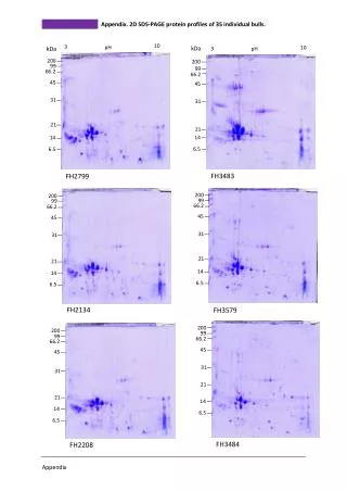
Experiment. Separation of main plasma protein by using SDS-PAGE. Prepared by Eman Alshehri. Objectives. Separation of plasma protein and determine its MWt. To learning some valuable skills in the field of separation of protein by SDS-PAGE. Identify the Varied uses of this technique.

E N D
Experiment Separation of main plasma protein by using SDS-PAGE Prepared by Eman Alshehri
Objectives • Separation of plasma protein and determine its MWt. • To learning some valuable skills in the field of separation of protein by SDS-PAGE. • Identify the Varied uses of this technique.
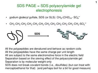
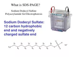
Introduction SDS-PAGE (Sodium dodecyle sulfate polyacrylamid gel electrophoresis) is method separates proteins based primarily on their molecular weight in electric field by uses a discontinuous polyacrylamide gel as a support medium and SDS to denature the proteins
Principle • Proteins are globular in secondary and tertiary structure due to disulfide bonds, hydrophobic interactions and hydrophilic interactions with their aqueous environment. Therefore, something must be done to break the secondary and tertiary structure of the proteins in the sample for accurate analysis of peptide size to occur. • Sodium dodecyl sulfate (SDS) is a detergent possessing both a hydrophobic end (the dodecyl group) and a hydrophilic end (the sulfate group).
The tertiary structure of most proteins often relies upon hydrophobic interactions at the core of the protein. The hydrophobic end of SDS breaks these interactions through interactions with the hydrophobic side chains of the amino acids. Similarly, a sulfate group can disrupt hydrogen bonding in secondary protein structure .
Disulfide bonds holding tertiary or quaternary structure together can be broken by using a reducing agent, such as beta-mercaptoethanol (BME). • Finally, heating the protein sample also aids the denaturation and unfolding process allowing chemicals like SDS and BME to interact with the protein. • In addition to denaturing the protein, SDS also serves an additional purpose. Because each protein is coated with SDS molecules and the charge. This means that when an electrical field is applied to the gel in buffer, each protein molecule will move toward the positive electrode. This allows the acrylamide to separate the proteins based on size
SDS disrupts the secondary, tertiary and quaternary structure of the protein to produce a linear polypeptide • chain coated with negatively charged SDS molecules. 1.4grams of SDS binds per gram of protein. • Mercaptoethanol assists the protein denaturation by reducing all disulfide bonds.
The Equipment SDS-PAGE set Power supply Boiling water for preparation sample.
Disruption buffer(sample buffer) • Sample Preparation • For best results, all samples should be in identical, low ionic strength buffers. • Mix 40 μl of each sample with 10 μl of disruption buffer. • Heat in a boiling water bath in for 2 min. • in most cases,brief boiling 3 min improves denaturation, but it may also cause the protein to precipitate.
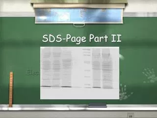
SDS PAGE Due to high density of binding of SDS to proteins, the ratio size/charge is nearly the same for many SDS denatured proteins. Hence proteins are separated only by length of their polypeptide chains (but not by differences in charge). Great separation. Allows estimation of the size of polypeptide chains
PAGE Separate native proteins by size –
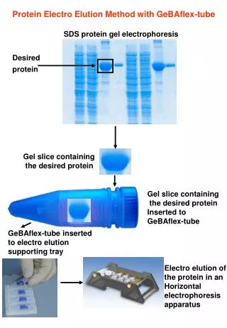
Protein visualization on gels • Immediately after electrophoresis proteins in the gels are often stained by Coomassie Blue dye.Stain the gel for 1 hour, agitate it slowly on a shaker. • Destain the gel in a destaining solution a few times until protein bands are visualised.