Download
1 / 24
240 likes | 403 Vues
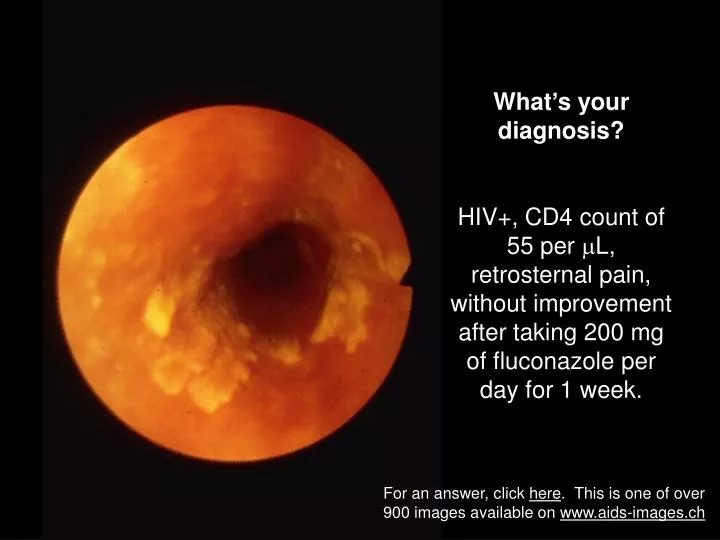
What’s your diagnosis? HIV+, CD4 count of 55 per L, retrosternal pain, without improvement after taking 200 mg of fluconazole per day for 1 week. For an answer, click here . This is one of over 900 images available on www.aids-images.ch. Answer:

E N D
What’s your diagnosis? HIV+, CD4 count of 55 per L, retrosternal pain, without improvement after taking 200 mg of fluconazole per day for 1 week. For an answer, click here. This is one of over 900 images available on www.aids-images.ch
Answer: Longitudinal ulcers in the lower esophagus suggest CMV esophagitis, confirmed by biopsy The prominent eosinophilic nucleolus in the submucosa of the esophagus is evidence for CMV infection This is one of over 900 images available on www.aids-images.ch
Sept 3, 2006 November 15, 2006 What’s your diagnosis? CD4 count of 184, fever, and weight loss For an answer, click here. This is one of over 900 images available on www.aids-images.ch
Sept 3, 2006 November 15, 2006 Answer: A partly calcified mass superior to the right kidney is seen on September 3, with increase in size on November 15. Remarkably high EBV viremia (7.1 * 106 per ml). The main differential diagnoses were lymphoma and tuberculosis. Needle biopsy yielded malignant cells which were CD20+, IRF4+, and Bcl-2+, but CD10 and Bcl-6 negative. Final diagnosis: malignant large-cell B type lymphoma This is one of over 900 images available on www.aids-images.ch
What’s your diagnosis? • 26 yo African woman with abdominal pain and suspected extrauterine pregnancy • HIV-test was +, CD4 count 18/L For an answer, click here. This is one of over 900 images available on www.aids-images.ch
Answer • Abdominal tuberculosis • CT shows extensive lymphadenopathy (arrows) • Laparoscopy revealed enlarged lymph nodes with acid-fast bacteria. Culture: M. tuberculosis • HIV-test was +, CD4 count 18/L This is one of over 900 images available on www.aids-images.ch
What’s your diagnosis? A 35 year-old patient with a CD4 count of 35, never treated for HIV. His wife reports that he forgets things and stumbles. (Left): MRI scan without gadolinium (Right): A T1-weighted image after gadolinium enhancement. For an answer, click here. This is one of over 900 images available on www.aids-images.ch
Answer: Probable AIDS-related dementia, a.k.a. HIV encephalitis, or AIDS dementia complex. (Left): Marked atrophy is evident: enlarged ventricles, prominent sulci (Right): The T1-weighted image after gadolinium enhancement shows diffuse, low-grade uptake of gadolinium in the white matter This is one of over 900 images available on www.aids-images.ch
What’s your diagnosis? This patient has established Castleman’s disease since October 2005. After chemotherapy he entered remission, but in October 2006, he had weight loss, fever, and precordial pain. Shown here is a frame from a 2-dimensional echocardiogram. For an answer, click here. This is one of over 900 images available on www.aids-images.ch
Answer: Pericardial effusion due to body-cavity lymphoma The echocardiogram shows an echo-free space surrounding the heart. Pericardiocentesis yielded an exudate with malignant plasmacytoid cells (see insert) staining for CD38, but negative for CD20 This is one of over 900 images available on www.aids-images.ch
What’s your diagnosis?An extremely immune suppressed patient died after a 7 month illness with malabsorption, loss of weight, fever, and finally liver and kidney failure. Biopsies of liver and duodenum showed massive infiltration by acid-fast bacteria, but cultures remained negative. Shown here: acid-fast stain of duodenum at autopsy. For an answer, click here. This is one of over 900 images available on www.aids-images.ch
Answer: Infection with Mycobacterium genavense, which clinically resembles infection with M. avium, except that conventional cultures remain negative. See N. Engl. J. Med. 323:109-13, and Lancet 1992 340:76-80 This is one of over 900 images available on www.aids-images.ch
What’s your diagnosis? Lesions gradually increased over several months.The patient is immune suppressed (CD4 count below 100) but otherwise asymptomatic. For an answer, click here. This is one of over 900 images available on www.aids-images.ch
Answer? Molluscum contagiosum. This is a benign viral infection, seen frequently in healthy children. In the presence of immune suppression, lesions may become more abundant. This is one of over 900 images available on www.aids-images.ch
“Crushed ping-pong balls” – that’s what this is supposed to resemble. What pathogen are we talking about? For an answer, click here. This is one of over 900 images available on www.aids-images.ch
Immunoperoxidase www.abcam.com Silver stain (« crushed ping-pong balls ») Giemsa Answer: There are many ways to color a pneumocyst Fungi-flor ® Grocott stain This is one of over 900 images available on www.aids-images.ch
What’s your diagnosis? An extremely immune suppressed patient presented with fever and heel pain. For an answer, click here. This is one of over 900 images available on www.aids-images.ch
Answer: Bacillary angiomatosis The diagnosis was established by biopsy (right, Whartin-Starry stain, and electron microscopy). Bartonella hensela shows up black rod-like or coccobacillary elements. Definite diagnosis relies on amplification and sequencing of 16SrDNA, as well as serology. This is one of over 900 images available on www.aids-images.ch
What’s your diagnosis? The symptoms were fever, chest pain, and dyspnea For an answer, click here. This is one of over 900 images available on www.aids-images.ch
Answer: Pneumocystis pneumonia (note interstitial infiltrate of the left lung) with spontaneous pneumothorax. Lung cysts may rupture spontaneously. Although spectacularly evident in the CT scan (right, from another patient), cysts are often difficult to see in conventional Xrays. This is one of over 900 images available on www.aids-images.ch
Lung involvement is often asymptomatic. Cryptococci are occasionally seen in fine needle aspirates of a coin lesion (left) or broncho-alveolar lavage samples (right) What’s your diagnosis? These micro-organisms were seen in broncho-alveolar lavage fluid. The symptoms were pulmonary infiltrates, fever, and headache. For an answer, click here. This is one of over 900 images available on www.aids-images.ch www.aids-images.ch
Lung involvement is often asymptomatic. Cryptococci are occasionally seen in fine needle aspirates of a coin lesion (left) or broncho-alveolar lavage samples (right) Answer: Cryptococcus neoformans, Papnicalaou stain. More usually, cryptococci are seen in the cerebrospinal fluid (right), using “india ink” staining. Budding forms are typical (arrow) This is one of over 900 images available on www.aids-images.ch www.aids-images.ch
What’s your diagnosis? For an answer, click here. This is one of over 900 images available on www.aids-images.ch
Answer: Advanced Kaposi’s sarcoma with bilateral leg edema, see detail (right) Photo credit: R. Lüthy This is one of over 900 images available on www.aids-images.ch