Download
1 / 82
880 likes | 1.36k Vues
Receptors. Seminar No. 9. Q. 1. Q. 2. A. 2. Allosteric protein (in membrane or cytosol) It has two domains: ligand-binding domain (with binding site for signal molecule) – changes conformations of receptor

E N D
Receptors Seminar No. 9
A. 2 • Allosteric protein (in membrane or cytosol) • It has two domains: • ligand-binding domain (with binding site for signal molecule) – changes conformations of receptor • effector domain – starts biological response to ligand (production of second messenger etc.)
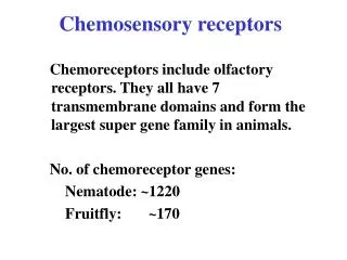
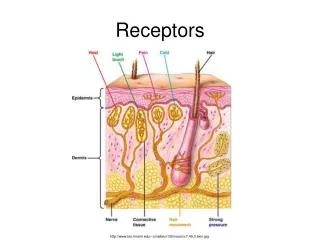
A. 4 • signal molecule(ligand) - carries specific information into cell • has extremely low concentration in blood (10-9 – 10-15 mol/l) • binds to corresponding receptor • signal molecule is usually quickly inactivated • agonist – ligand which after binding to receptor transduces signal • antagonist – ligand which after binding to receptor blocks signal transduction no biological response
A. 5 The second messenger transfers information to other intracellular systems and then is quickly inactivated Amplification of signal: 1 signal molecule 10 000-100 000 molecules of second messenger
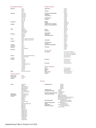
Factorsinvolved in biological action of hormones Hormone synthesis regulation effects Hormone storage feed back Hormonesecretion secretion impuls Transport in ECF metabolism inactivation Receptorin target cell cell condition excretion Biological response
A. 6 Examples of second messengers • Hydrophilic – cAMP, IP3 • Lipophilic – diacylglycerol (DAG) • Inorganic – Ca2+, NO
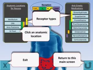
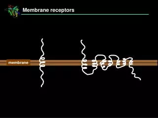
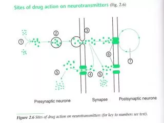
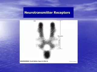
Two types of receptors: membrane and intracellular Three types of membrane receptors Ion channels in synapses, activated by neurotransmitters, very quick response Receptors activating G-proteins stimulate or inhibit adenylate cyclase /phospholipase C Receptors with enzyme activity guanylate cyclase - atrial natriuretic factors tyrosine kinase - insulin
Q. 11 Acetylcholine formation / inactivation
Acetylcholine formation acetyl-CoA choline acetylcholine food or synthesis from serine Inactivation ?
Acetylcholine inactivation acetylcholinesterase Acetylcholine + H2O Choline + acetic acid
GABA formation glutamate Inactivation ?
GABA inactivation GABA succinate semialdehyde oxid. deamination - NH3 oxidation succinate CAC
A. 12 + 13 • Excitatory neurotransmiters open cationic channels depolarization (more positive membrane potential) • Inhibitory neurotransmiters openanionic channels hyperpolarization(more negative potential)
Nicotinic acetylcholine receptor • transmembrane protein = channel for Na+ and K+ • heteropentamer (α2βγδ) • α-subunits have two binding sites for acetylcholine (ACH) • nicotine (= xenobiotic) is agonist of this receptor
Q. 15 – Four events on postsynaptic membrane and corresponding changes of membrane potential 2 1 3 4 1 2 3 4
A. 15 Four events on postsynaptic membrane • ACH binds to receptor channel opens influx of Na+and efflux of K+ membr. potential changes (-60 -40 mV) • partial depolarization of membrane opens voltage-dependent Na+-channel further influx of Na+ depolarization of postsyn. membrane ( +20 mV) • this depolarization opens K+-channel (volt. dep.) efflux of K+ membrane potential returns to normal value (-60 mV) = repolarization • Na+,K+-ATPase gets ion distribution to normal state (Na+ OUT, K+ IN)
GABA receptor • channel for chloride ion (Cl-) • has the binding site for GABA channel opens Cl- ions get into cell hyperpolarization ( -80 mV) decrease of excitability • benzodiazepines and barbiturates (synthetic substances) have similar effects like GABA, they are used as anxiolytics and/or sedatives • endozepines – endogenous peptides have opposite effects, close the channel (are responsible for anxiety feelings)
Diazepine Benzo[f]diazepine diazepine is a seven-membered unsaturated heterocycle with two nitrogen heteroatoms in the positions 1,4
Benzodiazepines structural modifications lead to different pharmacological effects
Barbiturates allobarbital: R1 = R2 = -CH2-CH=CH2 derivates of 2,4,6-trioxoperhydropyrimidine
Receptors with adenylate cyclase system(Scheme on p. 4) Describe the pathway of signal
G-Protein linked receptors • extracellular part of receptor has a binding site for hormone • intracellular part has a binding site for G-protein • G-proteins are heterotrimers (αβγ) • in resting state, α-unit has GDP attached • after binding hormone (α-GDP)βγ makes complex with receptor GDP is phosphorylated to GTP • activated G-trimer dissociates: (α-GTP)βγ α-GTP + βγ • α-GTP interacts with effector (enzyme) activated/inhibited enzyme second messenger (↑ or ↓)
What reaction is catalyzed by adenylate (adenylyl) cyclase? ATP cAMP What is adenylyl ?
Adenylyl cyclase reaction - diphosphate ATP cAMP cAMP = cyclic 3’,5’-adenosine monophosphate
AMP is called also adenylic acid adenylate is anion adenylyl is acyl
Adenylate (adenylyl) cyclase (AC) • membrane bound receptor • catalyzes reaction: ATP cAMP + PPi • Gs protein stimulates AC conc. of cAMP ↑ • Gi protein inhibits AC conc. of cAMP ↓
A. 19 • Protein kinase – phosphorylation by ATP Protein-OH + ATP Protein-O-P + ADP • Protein phosphatase – hydrolysis of phosphate ester Protein-O-P + H2O Protein-O-H + Pi
General scheme of phosphorylation substrate (protein) Which AA are phosphorylated? protein kinase phosphorylated protein (2-)
write structural formulas Three amino acids have a hydroxyl group in the side chain • Serine (3 C, primary alcohol hydroxyl) • Threonine (4 C, secondary alcohol hydroxyl) • Tyrosine (3 + 6 C, phenolic hydroxyl)
Phosphatidyl inositol system(Scheme on p. 4) Describe the pathway of signal
The structure of PIP2 describe the structure
Q. What is the source of inositol in human body?
The origin of inositol Exogenous source: any plant food (inositol hexaphosphate = phytic acid) Endogenous source: glucose-6-P (side path of metabolism)