Download

1 / 15
420 likes | 1.37k Vues
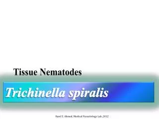
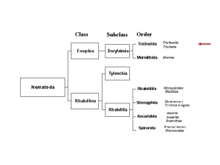
Trichinella spiralis. Presented by: Lauren Hannan and Chloe Jensen. Taxonomy. Kingdom: Animalia Phylum: Nematoda Class: Adenophorea Order: Trichurida Family: Trichinellidae Genus: Trichinella Species: T. spiralis. Introduction.

E N D
Trichinellaspiralis Presented by: Lauren Hannan and Chloe Jensen
Taxonomy • Kingdom: Animalia • Phylum: Nematoda • Class: Adenophorea • Order: Trichurida • Family: Trichinellidae • Genus: Trichinella • Species: T. spiralis
Introduction • Trichinella spp. is the smallest nematode parasite of humans, which has the most unusual life cycle, and is one of the most widespread and clinically important parasites in the world • Worlds largest intracellular parasite • T. spiralis is actually several strains – 8 “sibling species” are recognized • No morphological differences among the different kinds of Trichinella • Geographic distribution: worldwide
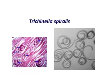
Morphology • Males are about 1.4 mm – 1.6 mm long, and are flat anteriorly and posteriorly • Have a large copulatorypseudobursa on each side • Females are about twice the size as males • Vulva is found near the esophagus • The single uterus of the female is filled with developing eggs in the posterior portion, while the anterior portion contains the fully developed juveniles.
Hosts • Definitive • Carnivorous and omnivorous animals, such as pigs or bears • Intermediate hosts • Primarily rodents • Accidental hosts • Humans
Life cycle • Trichinellosis is acquired by ingesting meat containing cysts (encysted larvae) of Trichinella • Once larvae enter the stomach and are exposed to digestive enzymes such as gastric acid and pepsin, the larvae burst out of the cysts and invade the small intestine, where they develop into adults • Live for about 4 weeks in the small intestine • After 1 week, females release larvae that migrate to the striated muscles and encyst – alter gene expression of the host cell! • Complete encystment takes about 4-5 weeks • Encysted larvae can remain viable for several years
Life cycle continued • Life cycle traditionally considered to be two epidemiologically distinct types: a domestic (involving pigs and rats, around human habitation), and a sylvatic (involving wild animals) • Rodents are primarily responsible for maintaining the endemicity of this infection • Carnivorous/omnivorous animals, such as pigs or bears, feed on infected rodents or meat from other animals • Humans become accidentally infected when eating improperly processed meat of these animals (or when eating food contaminated with such meat)
Pathogenesis • Causes Trichinosis • Three stages of pathogenesis: • Stage 1: penetration of adult females into the mucosa: • 12 hours – 2 days after infection • Low grade infection • Worms migration in intestinal epithelium causes: • Traumatic damage to the host tissues • Inflammation • Nausea, vomiting, diarrhea, • Sweating • Respiratory difficulties • Red skin blotches • Stage ends with facial edema and fever
Pathogenesis Continued • Stage 2: migration of juveniles: • Damages blood vessels, creating: • Localized edema • Pneumonia, pleurisy • Encephalitis, meningitis, nephritis, peritonitis • Deafness, brain/eye damage • Sublingual hemorrhage • Death from myocarditis may occur • Don’t stay in the heart, migrate through causing necrosis and infiltration of leukocytes
Pathogenesis Continued • Stage 3: Juvenile penetration of muscle fibers: • Symptoms are varied and vague • Intense muscle pain • Difficulty breathing/swallowing • Masseter muscle swelling • Weakening pulse and blood pressure, heart damage • Various nervous disorders • Death by heart failure, respiratory complications, and kidney malfunction • Heavy infection causes: • Reduced heart muscle contraction • Reduced stress, work, and power output
Diagnosis • Most cases go undetected • Routine exams rarely detect juveniles • Muscle biopsy is the most accurate form of diagnosis • Digestion of the muscle in artificial gastric enzymes for several hours
Treatment • No treatment • Just relieve symptoms • Analgesics and corticosteroids • Purges during beginning symptoms can dislodge worms • Thiabendazole • Effective in animals • Human trials have been variable
Control • Cook pork and meats properly and freeze unused meat • Watch out for “backyard butchering” • Don’t used uncooked garbage for pig food • Keep pig pens clean • Proper hygiene and sanitation
Review • What is the geographic range of this parasite? • What kind of hosts are humans? • T/F: Males are larger than females • After the larvae leave the small intestine, where do they migrate to? How long can they survive there? • What is the treatment method? • Name a control method • What is the most effective diagnosis method? • How does death typically occur?