Download
1 / 27
270 likes | 431 Vues
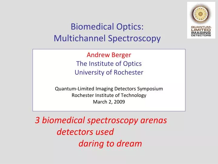
Andrew Berger The Institute of Optics University of Rochester Quantum-Limited Imaging Detectors Symposium Rochester Institute of Technology March 2, 2009. Biomedical Optics: Multichannel Spectroscopy. 3 biomedical spectroscopy arenas detectors used daring to dream.

E N D
Andrew Berger The Institute of Optics University of Rochester Quantum-Limited Imaging Detectors Symposium Rochester Institute of Technology March 2, 2009 Biomedical Optics: Multichannel Spectroscopy 3 biomedical spectroscopy arenas detectors used daring to dream
Biomedical Optics: Application Areas • diffuse photon propagation • fluorescence lifetime spectroscopy • Raman spectroscopy • barely imaging!!!
Where biomedical optics lives…. DNA biologicalwindow courtesy V. Venugopalan, http://www.osa.org/meetings/archives/2004/BIOMED/program/#educ
Important near-IR absorbers 19 M water 32 mM HbO2 11mM Hb 0.3 g/cm3 fat
Near-infrared cerebral blood monitoring light in (690, 830 nm) light out
Decoded Wavelength Data l830 l690 Brain monitoring system layout 1-10 kHz modulationfor wavelength encoding Analog Out DAQ Card 830 nm Source 1 High Speed DAQ Card for demultiplexing 690 nm 830 nm Avalanche photodiodes Source 2 near 690 nm near far far far far far far Sample
Typical detector for NIRS work • Hamamatsu silicon avalanche photodiode modules • Frequency rolloff in low MHz to GHz • Spectral response out to 1000 nm
Time-resolved measurements pulse at t=0 remitted light at t > 0 r absorption and scattering
Hand-Held Optical Breast Scanner Pham, TH., et al. Review of Scientific Instruments, 71 , 1 – 14, (2000). Bevilacqua, F., et al. Applied Optics, 39, 6498-6507, (2000). Jakobowski et al., J. Biomed. Opt., 9(1), 230-238 (2004). (courtesy F. Bevilacqua)
Heavily multiplexed systems! B. W. Pogueet al, Opt. Express 1, 391-403 (1997),http://www.opticsexpress.org/abstract.cfm?URI=OPEX-1-13-391
Diffuse propagation: goals, requirements • Distinguish benign from malignant tumor tissue • Map blood activity (hemodynamics) within brain • Sense deep within tissue (cm) • Record at many locations • Record at many wavelengths • Time resolution to few psec
Area #2: Fluorescence lifetime spectroscopy Once again, psec-nsec timescale!
Fluorescence lifetime spectroscopy brain tissue Butte et al., “Diagnosis of meningioma by time-resolved fluorescence spectroscopy,” Journal of Biomedical Optics 10(6), 064026 (November/December 2005).
Instrumentation for temporal fluorescence Fang et al.
Fluorescence lifetime: goals, requirements • Distinguish benign from malignant tumor tissue • Record at many wavelengths • Time resolution required to few psec • Desirable to record at many locations (imaging)
Area #3: Raman spectroscopy incident photonwith energy E molecule
Raman spectroscopy incident photonwith energy E molecule gains energy DE scattered photon has energy E -DE todetector
Raman spectrum of immune cell aromatic amino acids 1340 RNA bases 1092 1259 720 phenylalanine 902 intensity (arb. units) 853 667 813 1580 1005 1651 1457 1127 619 1211 amideIII 783 adenine tyrosine guanine phenylalanine C-H 2 def. amide I cytosine, uracil C-N, C-C str. Raman shift (cm-1)
Detectors for Raman spectroscopy • Thermoelectrically-cooledCCD array detectors • Sensitive out to ~1150 nm,limited by Si bandgap • 25 micron square pixels • typical dimensions, 256 x 1024 pixels Princeton Instruments PIXIS CCD
Raman spectroscopy: goals, requirements • Distinguish one cell type/state from another • Quantify chemical levels in biofluids (e.g. blood) • Yes, distinguish cancer from non-cancer • Record at many wavelengths • Long acquisition times (sec-minutes) • Necessaryto wavelength-tune down the fluorescence • Desirableto time-gate away the fluorescence (intensified CCD or more exotic gating)
Diffuse photons Fluorescencelifetime Raman Benefits of QLIDs for biomedical optics psec temporalresolution spectralresolution spectralrange thousandsof pixels [noise...]
Summary • biomedical spectroscopy: characterize tissue, biofluids, cells • frequently in near-IR • multiple factors driving sub-nsec time resolution • many-many-channel sensing: a game-changer • get past the Si bandgap cutoff • spectral resolution at each pixel: good for diffuse spectroscopy Questions?