Download
1 / 6
60 likes | 139 Vues
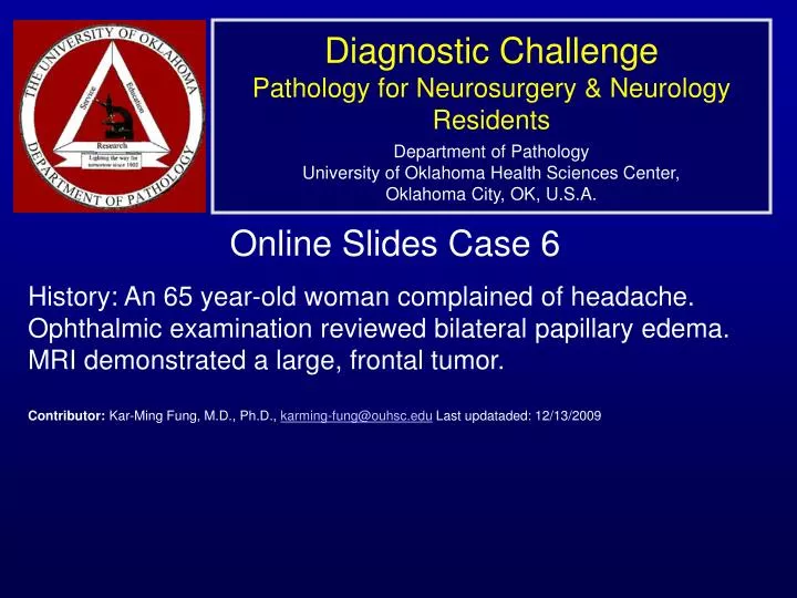
Diagnostic Challenge Pathology for Neurosurgery & Neurology Residents Department of Pathology University of Oklahoma Health Sciences Center, Oklahoma City, OK, U.S.A. Online Slides Case 6

E N D
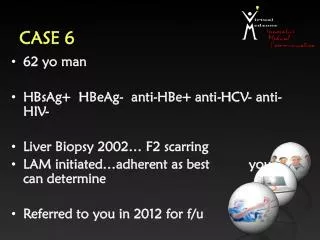
Diagnostic ChallengePathology for Neurosurgery & Neurology ResidentsDepartment of PathologyUniversity of Oklahoma Health Sciences Center,Oklahoma City, OK, U.S.A. Online Slides Case 6 History: An 65 year-old woman complained of headache. Ophthalmic examination reviewed bilateral papillary edema. MRI demonstrated a large, frontal tumor. Contributor: Kar-Ming Fung, M.D., Ph.D., karming-fung@ouhsc.edu Last updataded: 12/13/2009
T1 Contrast FLAIR B T2 A C
ß ß ß A B
ß ß Þ C D
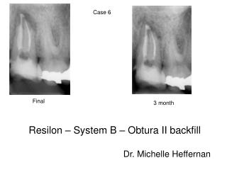
Diagnosis: Glioblastoma (WHO IV). • Discussion: • On the cytologic preparation, there are elongated cytoplasmic processes which is a tale telling feature of glioma (black arrow in anel A) and these cells have large, bizarre nuclei (white arrow in Panel A). • On permanent sections, the tumor is highly cellular and pseudopalisading necrosis is present at the corner (delimited by the two arrows in Panel C). Similar to the cytologic preparation, the nuclei are highly anaplastic and bizarre. • There are endothelial proliferation (white arrow in Panel C) although the proliferation looks incipient in this case. • These features are classic for glioblastoma (WHO grade IV).