Download
1 / 25
250 likes | 331 Vues
DNA damage, protein expression and migration of melanoma cells irradiated with proton beam. M. Elas 1 , S. Kędracka-Krok 1 , U. Jankowska 1 , Ł. Skalniak 1 , J. Jura 1 , E.Zuba-Surma 1 , K. Jasińska 1 , A. Pawlak 1 , U. Sowa 2 , P. Olko 2 , B. Romanowska-Dixon 3 , K. Urbańska 1

E N D
DNA damage, protein expression and migration of melanoma cells irradiated with proton beam M. Elas1, S. Kędracka-Krok1, U. Jankowska1, Ł. Skalniak1, J. Jura1, E.Zuba-Surma1, K. Jasińska1, A. Pawlak1, U. Sowa2, P. Olko2, B. Romanowska-Dixon3, K. Urbańska1 1Faculty of Biochemistry, Biophysics and Biotechnology, JU 2Institute of Nuclear Physics, PAS 3Department of Ophthalmology and Ophthalmic Oncology, Jagiellonian University Medical College
Methods BLM cells irradiated with 1-7 Gy of proton beam 58 MeV proton beam AIC-144 cyclotron at Institute of Nuclear Physics, Polish Academy of Sciences, Kraków LET <20 keV/µm dose rate 0.15 Gy/s
Dose-dependent slowing down of the proliferationrate 5 & 7 Gy lethal damage 1-3 Gy repair
Cell cycleredistribution: Increasein G2/M with dose, day 6 G0&G1/G2&M Increase in G2/M >2n present
DNA damage dose-dependent DNA damage 2 daysafterirradiation: no difference 5 daysafter: 11% more for 3 Gy, 14% for 5 Gy, 17% for 7 Gy =>delayedDNA damage
Caspases activity increases with time day 5 increase in caspases 3 and 7 activity with time
S91 S91 – day 9-10 S91 – day 5 3 Gy 3 Gy Apoptotic Cells Healthy Cells
Proteomics • 2DE and mass spectroscopy to identifyproteinsinfluenced by 3 Gy proton beamirradiation ctrl 3 Gy Kędracka-Krok et al., Plos One, 2014
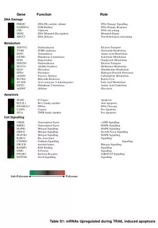
upregulated downregulated 13 up, 4 down > 1.5 x Nucleus VCP MVP STRAP FAB-2 Lamine A/C GAPDH Moesin Actinin 4 FAB-2 Vimentin Annexin 7 Lamine A/C Lamine B Cytoplasm STRAP MCM7 Annexin 7 MVP Caprin-1 PDCD6 VCP HSP70 P-body PDCD6 Caprin-1 TIM, GAPDH VCP Stress granule Mitochodrium Kędracka-Krok et al., Plos One, 2014
DNA repair, RNA regulation VCP MVP STRAP FAB-2 Lamine A/C GAPDH Nucleus Moesin Actinin 4 FAB-2 Vimentin Annexin 7 Lamine A/C Lamine B Cytoplasm STRAP MCM7 Annexin 7 MVP Caprin-1 PDCD6 VCP MVP (1.9 ), LamineA/C (1.8 ), GAPDH(2.4 ) – DNA repair VCP (1.9 ) – chromatinassociateddegradation MVP – PTEN translocationregulation MVP, VCP – transcriptionregulation STRAP (4.1 ) , FAB-2(1.9 ) – mRNAregulation Caprin-1 (1.9), PDCD6 (1.5 )– mRNAregulation HSP70 & G3BP1 (1.8 ) – stress & mRNA stability and stress granule formation PDCD6 P-body Caprin-1 TIM, GAPDH VCP Stress granule Mitochodrium Kędracka-Krok et al., Plos One, 2014
Cell survival VCP MVP STRAP FAB-2 Lamine A/C GAPDH Nucleus Moesin Actinin 4 FAB-2 Vimentin Annexin 7 Lamine A/C Lamine B Cytoplasm STRAP MCM7 Annexin 7 MVP Caprin-1 PDCD6 VCP STRAP (4.1 ) – survival - apoptosisbalance MCM7 (1.6 )– proliferationbalance Annexin 7(2.5 ) – proliferationinhibition MVP – survivalenhancement Caprin-1 (1.9) – proliferation PDCD6 (1.5 )– apoptosis, ubiquitinationregulation VCP (1.9 ) – aggregatesmanagement, lysosomaldegradation PDCD6 P-body Caprin-1 TIM, GAPDH VCP Stress granule Mitochodrium Kędracka-Krok et al., Plos One, 2014
Cell metabolism VCP MVP STRAP FAB-2 Lamine A/C GAPDH Nucleus Moesin Actinin 4 FAB-2 Vimentin Annexin 7 Lamine A/C Lamine B Cytoplasm STRAP MCM7 Annexin 7 MVP Caprin-1 PDCD6 VCP TIM (2.4 ), GAPDH(2.4 ) – glycolysis VCP(1.9 ) – mitochondria associateddegradation PDCD6 P-body Caprin-1 TIM, GAPDH VCP Stress granule Mitochodrium Kędracka-Krok et al., Plos One, 2014
Cytoskeleton and motility VCP MVP STRAP FAB-2 Lamine A/C GAPDH Nucleus Moesin Actinin 4 Fab-2 Vimentin Annexin 7 Lamine A/C Lamine B Cytoplasm STRAP MCM7 Annexin 7 MVP Caprin-1 PDCD6 VCP Moesin(2.4 ) – actinremodelling, motility Actinin 4 (1.9 ) – migration and metastasis FAB-2 (1.9 ) – migration, microtubuledestabilizer Vimentin (2.0, 2.1, 3.4, 1.6 ) –metastasis, EMT marker Annexin 7(2.5 ) – motility LamineA/C (1.6, 2,4, 1,5 ) – PI3K/AKT/PTEN, adhesion, motility Lamine B (1.4 ) - motility PDCD6 P-body Caprin-1 TIM, GAPDH VCP Stress granule Mitochodrium Kędracka-Krok et al., Plos One, 2014
Proteomics • 4 groups: • DNA repair & mRNAregulation • Survival and apoptosis • Glycolyticmetabolism • Cytoskeleton and migration • Signalingpathways: • p53 • TGFβ • PTEN • AKT • BAX, Bcl-2 Kędracka-Krok et al., Plos One, 2014
Interaction with tumor microenvironment • Heavy ionsshown to inhibitmetastasis (Takahashi, 2003; Tsuboi, 2005) • After proton beamirradiation: • strongly inhibited matrix metaloproteinase-2 activity in highly aggressive HT1080 human fibrosarcomacells in vitro, and significantly decreased the number of pulmonary metastasis in mouse osteosarcoma in vivo (Ogata et al. 2005). • the expression level or activity of molecules related to metastasis such as αVβ3, β1 integrin, and MMP-2 (Takahashi, 2003)
Angiogenesisgeneactivation Real-time PCR analysis usinghuman angiogenesis TaqMan® Array Plates Blm cells, 3 Gy, 48 hrculture Genes with 1.5 change vs controlshown
Migration properties of melanoma cellsafterprotonotherapy Blm, speed Omm1.3, speed Omm 1.3, distance Blm, distance
Inhibition of metastases of eye-implanted BHM melanoma 10 Gy Romanowska et al., ABP, 2013
Inhibition of metastases of eye-implanted BHM melanoma Rel mean diameter [mm] Time [days]
WYNIKI Inhibition of metastases of eye-implanted BHM melanoma x 4.4 p=0.0052 β-irradiation Romanowska et al., ABP, 2013
Conclusions • 1-3Gysublethal, 5-7 Gylethaldamage of BLM cells • Delayed DNA damage, leading to apoptosis, resulting from endogenous ROS generationdue to cellsignalling • Upregulation of proteinsinvolved in DNA repair and stress, apoptosis and survival, glycolyticmetabolism and migration and cytoskeleton • In the lattergroup, vimentin was heavilysuppressed, together with Annexin 7, whereasexpression of Moesin 7, Lamins A/C and B, Actinin 4 and FAB-2 wereincreased • Interaction with tumor microenvironment: Upregulation of angiogenicgenes, in vivo inhibition of metastases in BHM eye model
Obrazowanie mysich guzów: mechanizm odpowiedzi na radio- i fototerapię MRI, angiografia ToF MRI, perfuzja ASL
Dept of General Biochemistry Jolanta Jura Łukasz Skalniak Agnieszka Cierniak Institute of Nuclear Physics, PAS Paweł Olko Jan Swakoń Urszula Sowa Marta Ptaszkiewicz Dept PhysicalBiochemistry Sylwia Kędracka-Krok Urszula Jankowska Dept Cell Biology Ewa Zuba-Surma Marta Michalik Dept of Ophthalmology and Ophthalmic Oncology, Jagiellonian University Medical College Bożena Romanowska-Dixon Dept Biophysics Krystyna Urbańska Katarzyna Jasińska Małgorzata Szczygieł Martyna Krzykawska-Serda Michał Gonet Agnieszka Drzał
ODLEGŁE PRZERZUTY CZERNIAKA Przerzuty do płucsą inicjowane u części zwierząt (37%)gdy guz pierwotny zajmuje zaledwie 0,10 powierzchni PK(już po 2 dniach wzrostu guza)
WCZESNE I PÓŹNE EFEKTY RADIOTERAPII Całkowitej regresji guza po 10 Gy 125I (4,2,2,2) towarzyszy brak przerzutów w płucach u 50% leczonych chomików nawet po 70 dniach od enukleacji 29 –34 dni po ENU Średnica guza (mm) 100% zwierząt 70 dni po ENU Czas (dni) 50 % zwierząt (Sawow Aneta, 2001)