Download
1 / 20
240 likes | 579 Vues
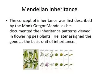
Mendelian inheritance in humans. Most traits in humans are due to the interaction of multiple genes and do not show a simple Mendelian pattern of inheritance. A few traits represent single-genes. Examples include sickle-cell anemia, cystic fibrosis, Tay-Sachs disease, and Huntington’s disease

E N D
Mendelian inheritance in humans • Most traits in humans are due to the interaction of multiple genes and do not show a simple Mendelian pattern of inheritance. • A few traits represent single-genes. Examples include sickle-cell anemia, cystic fibrosis, Tay-Sachs disease, and Huntington’s disease • Because we can not do breeding experiments on humans.
Three main categories of genetic disorders (1) Single-gene disorders (2) Chromosomal disorders (3) Complex disorders (multifactorial or polygenic) : hypertension, Diabetes mellitus Types of Single-Gene Disorders (Mendelian Disorders) (1) Autosomal Dominant Disorders (2) Autosomal Recessive Disorders (3) X-linked Disorders Single-Gene Disorders ( > 9,000 disorders recognized) (1) Victor A. McKusick’s “Mendelian Inheritance in Man” (12th edition, 1998) (2) Online version : Mendelian Inheritance in Man (OMIM) : continually updated. (3) >1,400 gene loci : mutations are associated with a clinically significant disorders (4) >90%: pediatric age range, <10%: after puberty, <1%: after the end of the reproductive period (5) 0.36% of live birth, 6-8% of hospitalized children (6) Every individual is a carrier of 4-8 deleterious genes (mostly recessive) 80-85% : familial, 15-20% : new mutations acquired de novo
Terminology Wild-type allele vs. Mutant type allele Mutation vs Polymorphism Genotype vs. Phenotype Genotype frequency, phenotype frequency, allelic frequency Homozygote, heterozygote (compound & double heterozygote), hemizygote
Anatomy of a pedigree Dizygotic & monozygotic twin Heterozygote Spontaneous abortion Pregnancy Multiple union Still birth Miscarriage No offspring
A vertical pattern of inheritance indicates a rare dominant trait Huntington’s disease: A rare dominant trait Assign the genotypes by working backward through the pedigree
Autosomal Dominant Disorders • Manifested in the heterozygote or homozygote state • Vertical inheritance: at least one parent of the index case is usually affected • Equal probability: both male and female can transmit the condition • Siblings have 50% chance for the recurrence • *New mutations in germ cells of parents normal parents but affected child • Transmission of new mutations depends on their effect on reproductive capability • Ex) Achondroplasia (short-limbed dwarfism) : reduced reproductive fitness • Thus, nearly all achondroplasias occurs by new mutations • ------------------------------------------------------------------------------------------------------------ • located on non-sex chromosomes • at least one parent is affected • does not skip generations • affected individuals are homozygous dominant or heterozygous • affects males & females • Achondroplasia, Huntington’s disease, Lactose intolerance, Polydactyly
Autosomal Recessive Disorders • Manifested in thehomozygote state (both alleles are mutants) • Horizontal inheritance: patrents are normal, but siblings show the disease • Siblings have 25% chance for the recurrence • Consanguineous marriage has a high recurrence risk for a rare disease • A certain mutant gene is common in population • Cystic fibrosis: White • Tay-Sacchs disease: Ashkenazi Jews or Central East Europe • Sickle cell anemia: Black • *Quasi-dominance: carrier X affected marriage: 50% offspring affected • ------------------------------------------------------------------------------------------------------------- • located on non-sex chromosomes • parents are carriers or are affected • affected individuals are homozygous recessive • affects males & females • Albinism, Cystic fibrosis, Phenylketonuria, Sickle cell disease
X-linked Disorders Affected male (hemizygous for X-liked genes) no sons are affected Carrier female 50% of sons are affected “No father to son transmission” is a hallmark of X-linked inheritance Hemophilia A (clotting factort VIII) Duchenne muscular dystrophy G6PD deficiency: red cell hemolysis in patients receiving certain drugs (Primaquine) If normal allele is inactivated in marrow cells drug-induced hemolysis X-linked disorder in female Random inactivation of X chromosome: Lyonization: Barr body If normal allele is inactivated in most cells full expression If normal allele is inactivated in only some of the cells partial expression
Dominance is not always complete Incomplete dominance : Phenotype severity is intermediate between homozygote and heterozygote Neither allele is dominant or recessive to the other Phenotypic ratios are same as genotypic ratios Codominance : F1 hybrids express phenotype of both parents equally Phenotypic ratios are same as genotypic ratios Histocompatibility, Blood group antigens
Sickle Cell Anemia Hb A/Hb A Hb A/Hb S Hb S/Hb S ------------------ -------------------------------- ------------------ Hg synthesis Normal Hb Normal & mutant Hb Mutant Hb Codominant Physiology Normal Mild anemia Anemia Incomplete dominant Clinical level A recessive trait
Factors Affecting Pedigree Patterns • Delayed Onset • Not all genetic disorders are congenital (congenital: “born with”) • Not all congenital disorders have a genetic basis • Huntington disease : average age of onest 35 years old • Familial adenomatous plyposis coli (FAP)
Factors Affecting Pedigree Patterns 2. Genetic Heterogeneity A number of phenotypes that are similar but are actually determined by different genotypes. Locus Heterozygosity: Similar disease phenotype caused by different genes Retinitis pigmentosa 3 X-linked, 12 autosomal dominant, & 5 autosomal recessive forms Ehlers-Danlos syndrome >10 different loci (X-linked, autosomal dominant or recessive) Childhood deafness Allelic heterozygosity: Different clinical phenotypes by different mutations at the same locus Different mutations in the RET gene Hirsch-sprung disease (defective colonic motility constipation) Multiple endocrine neoplasia type IIa and IIb
Locus Heterozygosity Allelic Heterozygosity Double heterozygote
Factors Affecting Pedigree Patterns 3. Pleiotrophism Single mutant gene may lead to many end effects : Sickle cell anemia 4. Codominance Histocompatibility, Blood group antigens 5. Reduced Penetrance “all or none” (% penetrance) normal persons can transmit the disease 6. Variable Expressivity expressed differentially Neurofibromatosis: brownish spots (café au lait spot) skin cancer
Four different situations in which one normal copy of the genes does not prevent disease 1. Haploinsufficiency Normal physiology requires more than 50% of fully active gene product 2. Dominant negative effect Abnormal protein causes an abnormal phenotype by interfering with normal protein function 3. Gain of function effect Mutant protein is enhanced or acquires a novel function through mutation 4. Predisposition to inherited cancers An inherited dysfunction of one allele results in pedigrees with inherited cancers