Download

1 / 62
620 likes | 730 Vues
Occupational Health II. By Dr. Muslim N. Saeed FAMCO dept./ Thi-Qar medical college April 8 th ,2019 4 th stage. Outlines. Definition of occupational disease and occupational environment. Interactions at work environment Types of occupational hazards and diseases

E N D
Occupational Health II By Dr. Muslim N. Saeed FAMCO dept./ Thi-Qar medical college April 8th ,2019 4th stage
Outlines Definition of occupational disease and occupational environment. Interactions at work environment Types of occupational hazards and diseases How to prevent occupational diseases
Definitions Occupational disease → Disease caused by or resulting from employment Occupational environment → sum of external conditions and influences which exist at work place and have an effect on the health of working population.
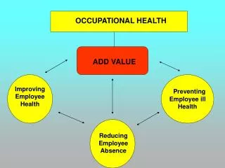
The aim of occupational health is to promote and maintain the highest degree of physical, mental and social well-being of workers in all occupations.
Basically, there are three types of interaction in a working environment: Man and physical ,chemical and biological agents. Man and machine. Man and man.
Man and Machine Accidents [lack of safety measures] Fatigue, backache and diseases of joints and muscles due to working for long hours in unphysiological postures.
Man and man Psychological stresses anxiety, industrial neurosis, depression, interpersonal conflicts.
Occupational Hazards Physical hazards Chemical hazards Biological hazards Mechanical hazards Psychological hazards
Occupational Diseases Diseases due to Physical agents Diseases due to Chemical agents Diseases due to Biological agents Diseases due to Mechanical agents Occupational cancers Occupational dermatosis Diseases due to Psychological origin
Lead poisoning is a medical condition caused by increased levels of the heavy metal lead in the body. Types of lead posisoning: 1. Acute lead poisoning from intense exposure to lead over short period of time 2. Chronic lead poisoning from repeated low-level exposure over long period of time. Chronic much more common than acute poisoning.
Lead uses and sources • Industrial uses: • Glass manufacture, ship building • Batteries, printing and potteries • In paints (in the past). • Plastic manufacturers • Rubber product manufacturers • Non-occupational sources: • Gasoline (thousands of tons of tetraethyl lead every year is exhausted from automobiles). • Drinking water from lead pipes. • Chewing lead paint on toys
Mode of absorption • Inhalation • Most common route 50-70%. Occur due to inhalation of fumes and dust of lead or its compounds. • Ingestion • Poisoning by ingestion is less common. • Small quantities of lead trapped in upper respiratory tract may be ingested .Lead may also be ingested in food or drink through contaminated hands. • Adults absorb about 6 - 10% of ingested lead. Fasting adults absorb more. • Children absorb muchmore lead (30-50% if well fed, and more, if fasting or malnourished). • Increased absorption if low Fe, Ca
Skin only in case of organic lead compounds (absorption through skin) Organic lead has greater affinity for CNS – therefore skin absorption may be SERIOUS. Lead Storage & Distribution • Rapid turnover soft tissue pool: • T1/2 30-40 days; blood, liver, kidney, CNS • Slow turnover skeletal pool: • T1/2 10-20 years
Distribution of Lead • 95% long bones. • 4% brain, liver, kidneys. • 1% blood. • Crosses placenta
Lead excretion • Renal (90%) and biliary (10%) • Maximum excretion is ~ 3.5µg/kg/day • If intake > 3.5 µg/kg/day accumulation will occur Lead metabolism and nutrition • Low dietary intake of vitamin D, vitamin C, and iron enhance absorption and retention of lead in the body. Body stores: • Body stores → 150 – 400 mg in adult. • Blood level → < 10 μg/100ml • Blood level ↑ to 30 - 40 μg/100ml → clinical symptoms
Health effects of lead exposure Organs affected by lead poisoning • CNS • Blood • Renal • GIT • Reproductive • Endocrine (including Bp) • Musculoskeletal
Clinical features of lead poisoning • I. Children Early symptom are nonspecific (anorexia, irritability, insomnia……),symptoms slowly intensify over time • Neurological symptoms • Developmental delay and loss of milestone especially language • Hearing loss • Peripheral neuropathy • Encephalopathy • Hematological→ hemolytic anemia • Renal → lead nephropathy • GIT → lead colic
II. In adults • The manifestations of lead poisoning can vary from individual to another. • Adults with severe lead poisoning (with blood lead levels generally above 80 μg/100ml ) can present with the following: • Abdominal pain ("lead colic"), constipation, joint pains, muscle aches, headache, hypertension anorexia, decreased libido, difficulty concentrating and deficits in short-term memory, anemia, nephropathy.
A "lead line," a bluish pigmentation seen at the gum-tooth line, is not a very sensitive finding, and is the result of a reaction of lead with dental plaque • A peripheral neuropathy that frequently manifests with extensor weakness or "wrist/ankle drop“ • Nephrotoxicity can occur in chronic poisoning
Diagnosis: • History of lead exposure • Clinical examination • LAB test • Blood lead level (BLL)The main tool in diagnosing and assessing the severity of lead poisoning . • The Free Erythrocyte Protoporphyrin (FEP) • EP increased when the amount of lead in the blood is high, with a delay of a few weeks . • EP levels in conjunction with blood lead levels can suggest the time period of exposure; if blood lead levels are high but EP is still normal, this finding suggests exposure was recent.
EP level alone is not sensitive enough to identify elevated blood lead levels below 35 μg/dL.Due to this and the fact that EP levels also increase in iron deficiency, use of this method for detecting lead exposure has decreased. • CBC : • basophilic stippling • Microcytic hypochromic blood picture • Renal function test (urea & creatinine )
Radiology (in children) • Radio-opaque lead flecks in the intestinal tract suggesting recent ingestion • Lead lines at the end of growing long bones (seen at end of metaphysis of long bones) in children.
Managment • Reduction or removal from lead exposure is the key first step in treating lead toxicity. • Chelation therapy • In most cases, removal from exposure is the only therapy needed. • Chelation is recommended for individuals with BLL >80 μg/100ml and those with levels between 60 -80 μg/100ml if they have lead-related symptoms. • Chelating agents : Promote lead excretion with urine • Ca-EDTA (Ca ethylene- diamine tetra- acetic acid) • DMSA (2,3-dimercaptosuccinic acid) • D-penicillamine. • Prevention of recurrence
Preventive measures • Periodic examination of the workers • Substitution • Isolation • Local exhaust ventilation • Personal protection and personal hygiene • Health education.
Occupational lung diseases (OLD) are caused by the inhalation of dusts, fumes, gases or vapors. Four main categories of OLD can be identified • Occupational asthma. • Chemical pnemonitis. • Pneumoconiosis [fibrotic and non-fibrotic]. • Granulomata.
Occupational asthma • Occupational asthma (OA) is characterized by airflow obstruction, airway hyperresponsiveness, and airway inflammation that results from a workplace stimulus. • Occupational asthma is one of the most common occupational lung diseases in developed countries. • 5 - 10% of adult-onset asthma is due to occupational exposure.
Occupations at risk • Animal handlers and veterinarians (animal proteins) • Bakers and millers (cereal grains) • Carpet makers (gums) • Cleaning staff (e.g. detergents , bleach) • Electronics workers (soldering resin) • Carpenters (wood dust) • Pharmaceutical workers (drugs, enzymes) • Seafood processors • Spray painters • Hair dressers • Health care workers (latex and chemicals)
Characteristics of Occupational Asthma • A workplace substance is aerosolized or vaporized • Patient has symptoms of asthma, but cough is the most common symptom. • Affects only some of those exposed • Onset often after symptom-free period of months to years • Improvement after removal from work early in course • Recurrence of symptoms on re-exposure
Clinical features • The latency period between exposure to stimuli and development of symptom varies among different stimuli. • Rhinitis and conjunctivitis may proceed or accompany asthma symptoms. • Cough, dyspnea ,chest tightness , wheezes. • The patient report increased symptoms while at work or within several hours of the completion of a shift, and definite improvement on weekends or during vacations.
Diagnosis • History and examination • Investigations • Pulmonary function test PFT • Skin test Managements: • Stop exposure • Treatment of occupational asthma is the same as for non-occupational asthma • The prognosis depends upon rapid diagnosis and prompt removal of the worker from further exposure • Most patients show incomplete resolution of asthma, even many years following cessation of exposure
Pneumoconiosis • The term “Pneumoconiosis” group of lung disorders which result from inhalation of dusts. (dust size 0.5 – 3 micron) • Many of these dusts give raise to fibrotic reaction in the lung with clinical symptoms. • Other group of dusts causes opacities on CXR, but no symptoms. These radiographic changes called Benign Pneumoconiosis
Classification • Benign pneumoconiosis • Siderosis (iron oxide lung) • Stannosis • Other Benign Pneumoconiosis • Fibrotic pneumoconiosis • Silicosis • Asbestosis • Coal Miner’s Pneumoconiosis (anthracosis)
Benign pneumoconiosis Siderosis (iron oxide lung) • The most common benign pneumoconiosis. • Result from inhalation of iron dust as iron oxide fume in iron and steel foundries during mining and during grinding and welding operation. Stannosis • Caused by deposition of tin in the lungs and it is less common than siderosis. The opacities are much denser with hilar L.N due to deposition of tin within them . Other Benign Pneumoconiosis: • Calcium, Barium (baritosis), Chromate, Zirconium and Cerium.
Fibrotic pneumoconiosis Silicosis • Disease produced due to inhalation of free silica or silicon dioxide (SiO2). • Occupational exposure: • works in mining (coal, gold, copper, silver , and lead) • Tunneling, stone cutting • Pottery and ceramic industry • Rock mining and metal grinding • The incidence of silicosis depends upon the chemical composition of the dust, size of the particles, duration of exposure and individual susceptibility.
The incubation period vary from few months up to 6 years, depending upon the above factors. Clinical features: Insidious onset, they are divided into arbitrary 3 stages which merge into one another. • 1st stage: irritant cough, dyspnea on exertion and chest pain. His working capacity is little affected or not affected. • 2nd stage: dyspnea with impaired ability to work. • 3rd stage: patient is totally incapacitated (signs of RHF). • Silicosis is associated with increased risk of Lung cancer&T.B
Prevention • Control of dust: • Substitution of the substance if possible. • Complete enclosure. • Isolation. • Wet procedure. • Regular cleaning (use of vacuum). • Personal protective measures. • Regular physical examination of the workers.
Asbestosis • Asbestosis specifically refers to the pneumoconiosis caused by inhalation of asbestos fibers. • The disease is characterized by slowly progressive, diffuse pulmonary fibrosis. • The spectrum of pulmonary disorders associated with asbestos exposure includes : • Asbestosis • Pleural disease (focal and diffuse benign pleural plaques) • Malignancies (non-small cell and small cell carcinoma of the lung as well as malignant mesothelioma)
Source of exposure • Mining and milling of the fibers • Industrial sources of asbestos (eg, work with textiles, cement, insulation, shipbuilding) • Non-occupational exposure to airborne asbestos (eg, regular exposure to soiled work clothes brought home by an asbestos worker, environmental exposure in the neighborhood of industrial sources….)
Clinical features • Symptoms are insidious with variable latent period. Dyspnea is the first symptom, which is frequently out of proportion to the clinical signs in the lungs • In advanced cases, there may be clubbing of fingers, cardiac distress , cyanosis , respiratory insufficiency and death. • The sputum shows “asbestos bodies” which are asbestos fibers coated with fibrin give rise to golden yellow or brown color
Prognosis→ once established; the disease is progressive even after removal of worker from contact. Prevention • Use of safer types of asbestos. • Substitution. • Rigorous dust control. • Regular physical examination of the workers (clinical ,CXR, lung function). • Health education and stop smoking. • Continuing research
Occupational skin diseases are very common, it responsible for 70% of occupational diseases. Classification of agents causing occupational skin diseases: • Mechanical agents : friction, pressure and trauma. • Physical agents : Heat, cold, humidity, light, ionizing radiation. • Chemical agents : both organic and inorganic chemicals. • Biological agents: Viral, bacterial and parasitic agents. • Plants and their products: leaves, fruits, dust, and other.
Mechanical agents • In the form of cuts, abrasions, repeated trauma → Calluses and blisters. • Callus is thickening of the skin in response to repeated trauma, friction or irritation. • Blisters result from acute trauma to the skin
Physical agents • Heat :Intertrigon,Erythemaabigne and burns • Cold → frost bite, chilblains. • Radiation → burns, dermatitis and cancer (BCC, SCC, melanoma). • Electricity→ Burns. Erythemaabigne: • Occurred in those exposed to furnaces, as cooks, glassblowers and kiln operators and long term exposure to a heating pad. • Pathogenesis: Long term exposure to radiant heat to the same area over time produces persistent vasodilatation of the dermal-subcutaneous blood vessels which leads to clinical changes.
Clinical Feature • The early stage is an asymptomatic reticulated pattern of the cutaneous blood vessels, which proceed to reticulated pigmentation called poikiloderma. • Areas of poikiloderma are prone to SCC and other types of skin cancers.
Cold induced skin injuries • Frost nip : The mildest cold-induced injury, characterized by localized paresthesia that resolve with rewarming. There is no permanent tissue damage. • Chilblains (Pernio) : is characterized by localized inflammatory lesions that result from acute or repetitive exposure to cold above the freezing point. Lesions are edematous, red, and may be very painful or pruritic. • Trench foot (immersion foot) • Involves the sympathetic nerves and vasculature of the feet. It resulted from prolonged exposure of the feet to the combination of dampness and cold.
Feet were red, edematous, numb or extremely painful, and often covered with hemorrhagic bullae. • Frostbite : • The most severe form of the localized cold-induced injuries. Frostbite results from the freezing of tissue. • The tissue destruction of frostbite is due to both immediate cold-induced cell death and the more gradual development of localized inflammatory processes and tissue ischemia
Chemical agents Contact Dermatitis: • Primary irritants contact dermatitis: Irritants can produce lesions by direct contact to the skin, and this depend on the concentration of substance and the duration of exposure. [e.g. acids, alkaline, dyes, solvents]. • Allergic contact dermatitis: Cause cell-mediated hyper- sensitivity reaction → Dermatitis.