Download
1 / 20
210 likes | 226 Vues
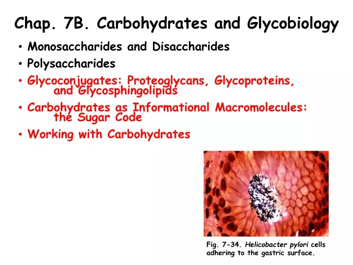
Chap. 7B. Carbohydrates and Glycobiology. Monosaccharides and Disaccharides Polysaccharides Glycoconjugates: Proteoglycans, Glycoproteins, and Glycosphingolipids Carbohydrates as Informational Macromolecules: the Sugar Code Working with Carbohydrates.

E N D
Chap. 7B. Carbohydrates and Glycobiology • Monosaccharides and Disaccharides • Polysaccharides • Glycoconjugates: Proteoglycans, Glycoproteins, and Glycosphingolipids • Carbohydrates as Informational Macromolecules: the Sugar Code • Working with Carbohydrates Fig. 7-34. Helicobacter pylori cells adhering to the gastric surface.
Intro. to Glycoconjugates In addition to their roles as energy storage and structural polymers, polysaccharides and oligosaccharides are information carriers. Some provide communication between cells and their extracellular surroundings; others label proteins for transport to and localization in specific organelles, or for destruction when the protein is malformed or superfluous; and others serve as recognition sites for extracellular signal molecules (growth factors, for example) or extracellular parasites (bacteria and viruses). On almost every eukaryotic cell, specific oligosaccharide chains attached to components of the plasma membrane form a carbohydrate layer (the glycocalyx), several nanometers thick, that serves as an information-rich surface that the cell presents to its surroundings. These oligosaccharides are central players in cell-cell recognition and adhesion, cell migration during development, blood clotting, the immune response, wound healing, and other cellular processes. In most of these cases the informational carbohydrate is covalently joined to a protein or a lipid to form a glycoconjugate, which is the biologically active molecule.
Glycoconjugate Classes Three types of glycoconjugates occur in nature (Fig. 7-24). Proteoglycans are macromolecules of the cell surface or extracellular matrix in which one or more sulfated glycosaminoglycan chains are joined covalently to a membrane protein or a secreted protein. Proteoglycans are major components of all ECMs. Glycoproteins have one or several oligosaccharides of varying complexity joined covalently to a protein. They are usually found on the outer face of the plasma membrane as part of the glycocalyx, in the ECM, or in the blood. The oligosaccharide portions of glycoproteins are very heterogeneous and are rich in structural information. Some cytosolic and nuclear proteins can be glycosylated as well. The glycosphingolipids are plasma membrane lipids in which the hydrophilic head groups are oligosaccharides. Both glycoproteins and glycosphingolipids are recognized and bound by carbohydrate-binding proteins called lectins.
Structure of Proteoglycans (I) Mammalian cells can produce 40 types of proteoglycans. These molecules act as tissue organizers, and they influence various cellular activities, such as growth factor adhesion and activation. The basic proteoglycan unit consists of a “core protein” to which glycosaminoglycan(s) are covalently attached. The point of attachment is a serine residue, to which the glycosaminoglycan is joined through a tetrasaccharide bridge (Fig. 7-25). The Ser residue is generally in the sequence -Ser-Gly-X-Gly-, where X is any amino acid. The xylose residue at the reducing end of the bridge is joined by its anomeric carbon to the hydroxyl of the Ser residue.
Structure of Proteoglycans (II) Many proteoglycans are secreted into the ECM, but some remain bound to the cell membrane of origin as integral membrane proteins. Two major families of membrane-bound heparan sulfate proteoglycans are described in Fig. 7-26a. The syndecans have a single transmembrane domain and an extracellular domain bearing three to five chains of heparan sulfate and in some cases chondroitan sulfate. Glypicans are attached to the membrane via a lipid anchor, which is a derivative of the membrane lipid phosphatidylinositol. Both syndecans and glypicans can be shed into the extracellular space after cleavage by a protease and a phospholipase, respectively, bound to the ECM. Proteoglycan shedding is involved in cell-cell recognition and adhesion, and in proliferation and differentiation of cells.
Structure of Proteoglycans (III) The glycosaminoglycan chains can bind to a variety of extracellular ligands and thereby modulate the ligands’ interaction with specific receptors on the cell surface. Glycosaminoglycans such as heparan sulfate contains domains (typically 3 to 8 disaccharides long) that differ from neighboring domains in sequence and in their ability to bind to specific proteins. Highly sulfated domains (called NS domains) alternate with domains having unmodified residues (NA domains, for N-acetylated domains) (Fig. 7-26b). The exact pattern of sulfation in the NS domains depends on the particular proteoglycan. Furthermore, the same core protein can display different heparan sulfate structures when synthesized in different cell types.
Examples of Proteoglycan Functions (I) Heparan sulfate molecules with precisely organized NS domains bind specifically to extracellular proteins and signaling molecules to alter their functions. In the first example (Fig. 7-27a), the binding of antithrombin (AT) to the heparan sulfate moiety of a proteoglycan in the ECM alters the conformation of AT so that it binds to the blood clotting factor Xa, preventing clotting. AT recognizes a specific sulfated NS domain in heparan sulfate.
Examples of Proteoglycan Functions (II) In another example of proteoglycan function (Fig. 7-27b), the binding of AT and thrombin to two adjacent NS domains of heparan sulfate chains in a proteoglycan brings them into close proximity, where they can bind to one another, and AT inhibits the activity of thrombin.
Proteoglycan Aggregates Some proteoglycans can form proteoglycan aggregates, enormous supramolecular assemblies of many glycosaminoglycan-decorated core proteins all bound to a single molecule of hyaluronan (Fig. 7-28). The core protein involved commonly is aggrecan (Mr 250,000). Aggrecan binds multiple chains of chondroitin sulfate and keratin sulfate via covalent linkages to serine residues. When a hundred or more of these decorated core proteins binds a single extended hyaluronan molecule the resulting proteoglycan aggregate has a mass of Mr >2 x 10⁸. Its size is comparable to a bacterial cell. Aggrecan interacts with collagen in the ECM of cartilage, contributing to the development, tensile strength, and resilience of this connective tissue.
Interactions Between Cells and the ECM The ECM is a strong and resilient meshwork containing proteoglycans in association with collagen, elastin, and fibronectin. Some of these proteins are multiadhesive, with a single protein having binding sites for several different matrix molecules. The ECM protein fibronectin, for example, has several separate domains that bind fibrin, heparan sulfate, collagen, and a family of plasma membrane proteins called integrins. Integrins mediate signaling between the cell interior and the ECM. A diagram showing just some of the interactions occurring between a cell and the surrounding ECM is shown in Fig. 7-29. These interactions not only anchor the cell in the ECM, but also provide paths that direct the migration of cells in developing tissue and convey information in both directions across the plasma membrane.
Structure of Glycoproteins Glycoproteins are carbohydrate-protein conjugates in which the glycans are smaller, branched, and more structurally diverse than the huge glycosaminoglycans present in proteoglycans. Two broad classes of glycoproteins occur in nature--the O-linked and the N-linked glycoproteins (Fig. 7-30). In O-linked glycoproteins, the carbohydrate is attached at its anomeric carbon through a glycosidic linkage to the -OH group of a Ser or Thr residue. Both a simple chain and a complex carbohydrate chain are shown in the figure. In an N-linked glycoprotein, the carbohydrate is attached via an N-glycosyl linkage to the amide nitrogen of an Asn residue of the protein. N-linked oligosaccharide chains are attached at Asn residues that are part of the consensus sequence -N-{P}-[ST]-, although not all such sequences in proteins are modified. There appears to be no specific consensus sequence for the attachment of O-linked oligosaccharides. Three types of N-linked oligosaccharide chains that are common in glycoproteins are shown in the figure. The mechanism of synthesis and attachment of these chains is covered in Chap. 27.
Functions of Glycoproteins About half of all proteins of mammals are glycosylated. Many of these are plasma membrane proteins and secretory proteins. The external surface of the plasma membrane has many membrane glycoproteins with vast types of covalently attached oligosaccharides of varying complexity. Examples of glycosylated secretory proteins include immunoglobulins (antibodies) and certain hormones such as follicle-stimulating hormone and luteinizing hormone. However, another class of glycoproteins found in the cytoplasm and nucleus carry only a single residue of N-acetylglucosamine attached in O-glycosidic linkage to the hydroxyl group of Ser side-chains. This modification is reversible and often occurs on the same Ser residues that are phosphorylated. The two modifications are mutually exclusive, and the modified protein exhibits different activity in the two modified states. The biological advantages of protein glycosylation include improving the solubility of proteins, providing address labels directing targeting in cells and between tissues, and protein folding and stabilization. At this time 18 different genetic diseases of protein glycosylation have been described in humans. Finally, the discipline of glycomics, the systematic characterization of all of the carbohydrate components of a given cell or tissue, offers insight into the normal patterns of glycosylation in cells and how they may be perturbed in diseases such as cancer.
Glycolipids and Lipopolysaccharides (I) In glycolipids and lipopolysaccharides, complex oligosaccharide chains are attached to membrane-anchored lipid moieties. The complex structure of the lipopolysaccharide found in the outer membrane of Salmonella typhimurium is shown in Fig. 7-31. These molecules, which are found in other Gram-negative bacteria such as E. coli, are recognized by the immune system of vertebrates and therefore are important determinants of the serotypes of bacterial strains. Serotypes are strains that are distinguished on the basis of antigenic properties. The fatty acid-containing lipid A portion of lipopolysaccharide is called endotoxin. Its toxicity to humans is responsible for the dangerously lowered blood pressure that occurs in toxic shock syndrome resulting from Gram-negative bacterial infections.
Glycolipids and Lipopolysaccharides (II) Plants and animals contain many types of glycolipids and glycosphingolipids. The gangliosides are glycosphingolipids containing complex oligosaccharide chains with sialic acid residues. These lipids are important for cell-cell recognition, and some serve as receptors for the entry of bacterial toxins such as cholera toxin into mammalian cells. Other glycosphingolipids, the globosides, contain oligosaccharide chains that serve as the blood group antigens. The structures of several glycosphingolipids are covered in Chap. 10.
Carbohydrates as Informational Molecules: The Sugar Code Glycobiology is the study of the structure and function of glycoconjugates. Cells use specific oligosaccharides to encode important information about intracellular targeting of proteins, cell-cell interactions, cell differentiation and tissue development, and extracellular signals. The oligosaccharides of glycoproteins and glycolipids are highly complex and diverse. Oligosaccharides of typical glycoproteins can contain a dozen or more monosaccharide residues in a variety of branched and linear structures, and in a variety of linkages. (See the N-linked oligosaccharides in the glycoprotein examples shown in Fig. 7-30). With the assumption that 20 different monosaccharide subunits are available for the construction of oligosaccharides, it can be calculated that many billions of different hexameric oligosaccharides, for example, are possible. This compares with 6.4 x 10⁷ (206) different hexapeptides possible from the 20 common amino acids, and 4,096 (46) different hexanucleotides with the four bases. The structural information potentially present in glycans thus actually surpasses that of nucleic acids for molecules of modest size. Each of the oligosaccharides attached to a glycoprotein for example, presents a unique three-dimensional structure--a word in the sugar code--that is readable by the proteins that interact with it.
Lectin Functions (I) Lectins, found in all organisms, are proteins that bind carbohydrates with high specificity and with moderate to high affinity. Lectins play roles in a wide variety of cell-cell recognition, signaling, and adhesion processes and in intracellular targeting of newly synthesized proteins. Some examples of these functions are covered in the next few slides. The first example covered concerns the role of the asialoglycoprotein receptor (a lectin) of the liver which is important in the clearance of many plasma glycoproteins from the circulation. Normally, many plasma glycoproteins are synthesized with oligosaccharide chains that terminate with N-acetylneuraminic acid (a sialic acid) (see figure). This sugar protects the glycoproteins initially from uptake and degradation by hepatocytes. However, during the lifetime of the glycoprotein in the bloodstream, its sialic acid residues are gradually removed by enzymes called neuraminidases (sialidases) of unknown origin. These asialo- versions of the original glycoproteins are then taken up by the liver and degraded. Note that a similar mechanism is apparently responsible for the removal of old erythrocytes from the bloodstream by the spleen.
Lectin Functions (II) Several animal viruses, including influenza virus, attach to their host cells though the interactions of viral lectins (e.g., influenza virus hemagglutinin protein) and oligosaccharides displayed on the host cell surface. After the virus has replicated inside the host cell, the newly synthesized viral particles bud out of the cell, wrapped in a portion of its plasma membrane. A viral sialidase trims the terminal sialic acid residues from the host cell’s oligosaccharides, releasing the viral particles from their interaction with the cell and preventing their aggregation with one another. Another round of infection can then begin. The antiviral drugs oseltamivir (Tamiflu) and zanamivir (Relenza) are used in the treatment of influenza. These drugs are sugar analogs that are structurally similar to sialic acid (N-acetylneuraminic acid) (Fig. 7-33a). They therefore can inhibit the viral sialidase and limit the release of viruses from the original infected cell.
Lectin Functions (III) The binding site on influenza neuraminidase (sialidase) for N-acetylneuraminic acid and the antiviral drug, oseltamivir (Tamiflu) is shown in Fig. 7-33 b-d. Tamiflu competitively inhibits the binding of terminal sialic acid residues of oligosaccharide chains to the neuraminidase. When Tamiflu binds to the active site it pushes away the side-chain of Glu276, making room for the binding of the inhibitor (Part c). Mutations in viral neuraminidase, however, which replace nearby His274 with a bulky Tyr residue, prevent the repositioning of Glu276, and Tamiflu binding to the active site (Part d).
Lectin Functions (IV) Some microbial pathogens have lectins that mediate bacterial adhesion to host cells or the entry of toxins into cells. For example, the gastric ulcer-causing bacterium, Helicobacter pylori, has a surface lectin that adheres to oligosaccharides on the surface of epithelial cells that line the inner surface of the stomach (Fig. 7-34). This allows the bacterium to colonize the stomach and cause associated ulceration. The H. pylori lectin actually binds to the Lewis b (Leb) antigen present in cell surface glycoproteins and glycolipids that defines the type O blood group. This observation helps explain the several-fold greater incidence of gastric ulcers in people of blood type O than in those of type A or B. Chemically synthesized analogs of the Leb antigen may prove useful in treating this type of ulcer.
Analysis of Glycoprotein Oligosaccharides An overview of some of the procedures used to analyze the total composition, linkages, and sequences of monosaccharides in oligosaccharide chains is presented in Fig. 7-38. The method starts with the removal of oligosaccharide chains from the glycoproteins that contain them by enzymes called endoglycosidases. It typically ends with the fine-structure analysis of the chains by NMR and mass spectrometry. Oligosaccharide structure analysis is more complicated than protein and nucleic acid analysis due to the branching and variety of linkages present in these molecules.