Download
1 / 55
560 likes | 695 Vues
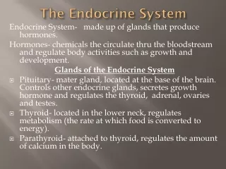
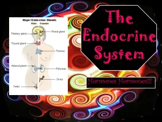
The Endocrine System. The Endocrine System: An Introduction. Hormones (from Greek Harmon – to excite) : are chemical signals that are secreted into body fluids (most often the blood) and communicate regulatory messages within the body

E N D
The Endocrine System: An Introduction • Hormones (from Greek Harmon – to excite) : are chemical signals that are secreted into body fluids (most often the blood) and communicate regulatory messages within the body • Target cells : Cells that are equipped to respond to hormones • Elicits specific responses
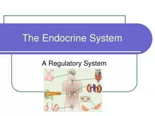
Our Primary Focus • Internal chemical signals • What they are • How they function • Their role in maintaining homeostasis • A dynamic, steady state in body functions
Regulation • Animals have 2 systems for regulation • Nervous System Regulation • Fast means of communication • Fast response • Endocrine System Regulation • Slower means of communication • Slower responses
The Endocrine System • All of an animals hormone secreting cells • Also called ductless glands • Secrete their chemical messengers directly into body fluids • Exocrine glands secrete into ducts that convey the message to the appropriate location • Mucus, sweat, digestive enzymes
Nervous system and Endocrine system similarities – more than you think! • Neruosecretory cells • Specialized nerve cells that secrete hormones • Both vertebrate and invertebrate • Eg. Epinephrine • Feedback • Positive or Negative
Figure 45.1 An example of how feedback regulation maintains homeostasis
Figure 45.2 Hormonal regulation of insect development (Layer 1) Molting is triggered by a hormone called ecdsone. It also favors the development of adult characteristics. It’s controlled by a second hormone, Brain Hormone (BH).
Figure 45.2 Hormonal regulation of insect development (Layer 2)
BH and ecdysone are balanced by juvenile hormone (JH). This hormone promotes the retention of larval characteristics. Synthetic versions of JH are being used as pesticides – lots of JH means not many adults. Will it work? Figure 45.2 Hormonal regulation of insect development (Layer 3)
Figure 45.0 A monarch butterfly just after emerging from its cocoon
1. A variety of local regulators affectneighboring target cells • Growth factors: proteins and polypeptides that stimulate cell proliferation • Example: nerve growth factor (NGF) affects certain embryonic cells, developing white blood cells, and other kinds of cells
Nitric oxide (NO) • Though a gas, NO is an important local regulator. • When secreted by neurons, it acts as a neurotransmitter. • When secreted by white blood cells, it kills bacteria and cancer cells. • And when secreted by endothelial cells, it dilates the walls of blood vessels.
Prostaglandins (PGs): modified fatty acids. • PGs secreted by the placenta stimulate uterine contractions during childbirth. • Other PGs play a role in inflammation and the blood flow to the lungs.
2. Most chemical signals bind to plasma-membrane proteins, initiating signal-transduction pathways. Fig. 45.3a
Different signal-transduction pathways in different cells can lead to different responses to the same signal. Fig. 45.4
Signal- transduction pathways allow for small amounts of a hormone to have a large effect. Fig.11.16
3. Steroid hormones, thyroid hormones, and some local regulators enter target cells and bind to intracellular receptors • Examples: estrogen, progesterone, vitamin D, NO. • Usually, the intracellular receptor activated by a hormone is a transcription factor. Fig. 45.3b
Introduction • Tropic hormonestarget other endocrineglands and areimportant tounderstandingchemicalcoordination. • Humans havenine endocrineglands. Fig. 45.5
4. The hypothalamus and pituitary integrate many functions of the vertebrate endocrine system • The hypothalamus integrates endocrine and nervous function. • Neurosecretory cells of the hypothalamus produce hormones. • Releasing hormones stimulate the anterior pituitary (adenohypophysis) to secrete hormones. • Inhibiting hormones prevent the anterior pituitary from secreting hormones.
The posteriorpituitary(neurohypo-physis)stores andsecretes hormonesproducedby thehypothalamus. Fig. 45.6a
Hormones manufactured by the hypothalamus and released by the posterior pituitary. • Oxytocin: a peptide. • Stimulates contraction of the uterus and mammary glands. • Secretion regulated by the nervous system. • Antidiuretic hormone (ADH): a peptide. • Promotes retention of water by the kidneys. • Secretion regulated by water/salt balance.
Anterior pituitary hormones. • Growth hormone (GH): a protein. • Stimulates growth and metabolism. • Secretion regulated by hypothalamic hormones. • Acts directly on tissues or acts via growth factors. • Gigantism: excessive GH during development. • Acromegaly: excessive GH production during adulthood. • Hypopituitary dwarfism: childhood GH deficiency.
Prolactin (PRL): a protein. • Stimulates milk production and secretion. • Secretion regulated by hypothalamic hormones. • Gonadotropins: glyocoproteins. • Follicle-stimulating hormone (FSH). • Stimulates production of sperm and ova. • Secretion regulated by hypothalamic hormones. • Luteinizing hormone (LH). • Stimulates ovaries and testes. • Secretion regulated by hypothalamic hormones.
Thyroid-stimulating hormone (TSH): a glycoprotein. • Stimulates thyroid gland. • Secretion regulated by thyroxine in blood. • Secretion regulated by hypothalamic hormones. • Adrenocorticotropic hormone (ACTH): a peptide • Stimulates adrenal cortex secretion of glucocorticoids • Secretion regulated by glucocorticoids and hypothalamic hormones.
Melanocyte-stimulating hormone (MSH): a peptide. • May play a role in fat metabolism. • Endorphins: peptides. • Inhibit pain perception. • Effects mimicked by heroin and other opiate drugs.
5. The pineal gland is involved inbiorhythms • The pineal gland is a small mass of tissue near the center of the mammalian brain. • The pineal gland secretes the hormone, melatonin, an amine. • Involved in biological rhythms associated with reproduction. • Secretion regulated by light/dark cycles.
6. Thyroid hormones function in develop-ment, bioenergetics, and homeostasis • The thyroid glandof mammals consistsof two lobes locatedon the ventral surfaceof the trachea. • Triiodothyronine (T3)and thyroxine (T4): amines. • Stimulates and maintainmetabolic processes. • Secretion regulated by TSHhormones. Fig. 45.8
Hyperthyroidismis the excessive secretion of thyroid hormones, exhibited by high body temperature, profuse sweating, weight loss, irritability, high blood pressure. • An insufficient amount of thyroid hormones is known as hypothyroidism. • Infants: cretinism. • Adults: weight gain, lethargy, cold intolerance. • Goiter: often associated with iodine deficiency. • Calcitonin: a peptide. • Lowers blood calcium levels. • Secretion regulated by calcium in blood.
Calcitonin: a peptide. • Lowers blood calcium levels. • Secretion regulated by calcium in blood.
7. Parathyroid hormone and calcitonin regulate blood calcium level • The four parathyroid glands are embedded in the surface of the thyroid gland.
They secrete parathyroid hormone (PTH), a peptide. • Raises blood calcium levels. • Secretion regulated by calcium in the blood. • Causes osteoclasts to break down bone, releasing Ca2+ into the blood. • Stimulates the kidneys to reabsorb Ca2+. • Stimulates kidneys to convert vitamin D to its active form. • PTH and calcitonin are antagonistic hormones. • Hypoparathyoidism: tetany.
A lack of PTH causes hypoparathyoidism, a tetany. • Calcium levels in the blood drop. • There are convulsive contractions of the skeletal muscles.
8. Endocrine tissues of the pancreas secrete insulin and glucagon, antagonistic hormones that regulate blood glucose • The pancreas has both endocrine and exocrine functions. • Exocrine function: secretion of bicarbonate ions and digestive enzymes. • Endocrine function: insulin and glucagon secreted by islets of Langerhans.
Insulin: a protein secreted by beta cells. • Lowers blood glucose levels. • Stimulates all body cells (except brain cells) to take up glucose. • Slows glycogenolysis. • Inhibits gluconeogenesis. • Secretion regulated by glucose in blood (negative feedback).
Hypoinsulinism: diabetes mellitus. • Hereditary factors and obesity play a role in its development. • High blood sugar levels – sugar excreted in the urine. • Symptoms: excessive urination and excessive thirst. • If severe: fat substitutes for glucose as major fuel source production of acidic metabolites life threatening lowering of blood pH.
Type I diabetes mellitus (insulin-dependent diabetes). • Autoimmune disorder. • Usually appears in childhood. • Treatment: insulin injections. • Type II diabetes mellitus (non-insulin-dependent diabetes). • Usually due to target cells having a decreased responsiveness to insulin. • Usually occurs after age 40 – risk increases with age. • Accounts for over 90% of diabetes cases.
Glucagon: a protein secreted by alpha cells. • Raises blood glucose levels. • Stimulates glyogenolysis in the liver and skeletal muscle. • Secretion regulated by glucose in blood (negative feedback).
9. The adrenal medulla and adrenal cortex help the body manage stress • The adrenal glands are located adjacent to the kidneys. • The adrenal cortex is the outer portion. • The adrenal medulla is the inner portion.
Adrenal medulla. • Developmentally and functionally related to the nervous system. • Epinephrine (adrenaline)and norepinephrine (noradrenaline). • Catecholamines: amines synthesized from tyrosine. • Secretion regulated by the nervous system in response to stress.
Epinephrine (adrenaline)and norepinephrine (noradrenaline). • Catecholamines: amines synthesized from tyrosine. • Secretion regulated by the nervous system in response to stress. • Raises blood glucose level and blood fatty acid level. • Increase metabolic activities. • Increases heart rate and stroke volume anddilates bronchioles. • Shunts blood away from skin, digestive organs, and kidneys, and increases blood flow to heart, brain, and skeletal muscle.
Adrenal cortex reacts to stress. • Secretion of corticosteroids is regulated by the nervous system in response to stress.
Glucocorticoids. • Raises blood glucose level. • Secretion regulated by ACTH (negative feedback). • Abnormally high doses are administered as medication to suppress the inflammation response.
Mineralocorticoids (example: aldosterone, which affects salt and water balance). • Promotes reabsorption of Na+ and excretion of K+ in kidneys. • Secretion regulated by K+ in blood.