Download
1 / 3

E N D
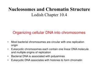
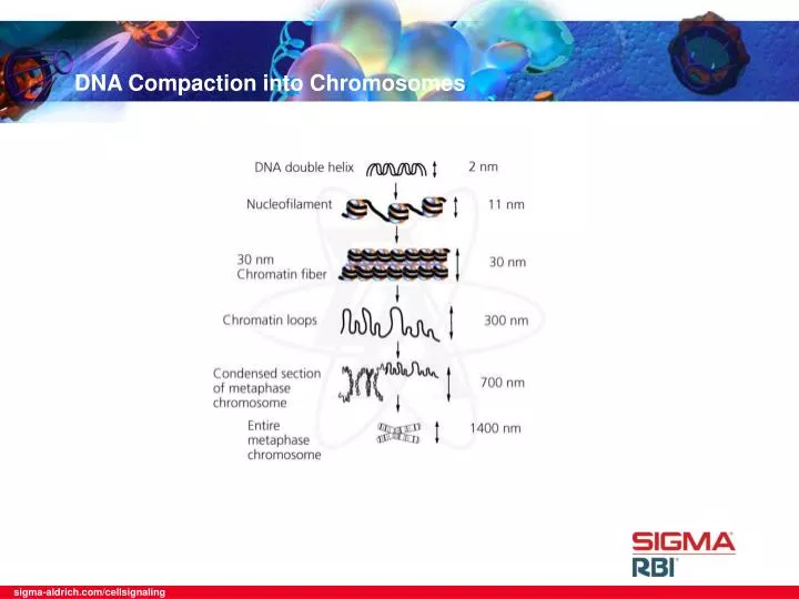
The structure of chromatin was first revealed by electron microscope analysis to be regularly spaced particles commonly described as “beads on a string”. Cross-linking studies demonstrated that the stoichiometry of DNA and histones in the nucleosome is 1:1 based on their mass. Taken together, these observations led to the hypothesis that the nucleosome was the fundamental unit of chromatin. The nucleosome is composed of a core particle and a linker region that joins adjacent core particles. The core particle structure is highly conserved between different species and is composed of 146 base pairs of DNA wrapped 1.7 turns around a protein octamer of two each of the core histones H3, H4, H2A and H2B. However, the length of the linker region varies markedly between different species and cell type. It is within this region that the variable linker histones are incorporated. The core histones, H3, H4, H2A and H2B, are small, basic proteins that are highly conserved throughout evolution. The most highly conserved region of these histones is their central domain that is structurally composed of the histone fold-helices separated by two loop regions. In contrast, the N-terminal tails of each core histone display more variability. The tail regions are rich in lysine and arginine residues which makes them extremely basic. This region is the site of numerous post-translational modifications that modify its charge and thereby alter DNA accessibility and protein:protein interactions with the nucleosome. DNA Compaction into Chromosomes
Assembly of DNA into chromatin involves a wide range of events, beginning with formation of the nucleosome. The next step in the process is the maturation step that requires ATP to establish regular spacing of the nucleosome cores to form the nucleofilament. During this step the histones are de-acetylated. Next, linker histones are incorporated and nucleofilament is folded into the 30 nm fiber. Further folding events lead to a high level of organization and specific domains within the nucleus. At each step, variation in the composition and activity of chromatin can be obtained by modifying its basic constituents and the activity of various stimulatory factors involved in its assembly and disassembly. Adapted from Textbook, “Molecular Biology of the Cell” Alberts B., Bray D., Lewis J., Raff M., Roberts K. and Watson J.D. DNA Compaction into Chromosomes