Download
1 / 1
10 likes | 131 Vues
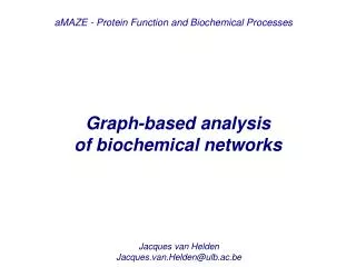
Figure 1 . PTS system diagram. Figure 4 . Simple membrane-bound decay reaction. (A m +B C) Notice the different decay rate at the membrane (x=1), and the horizontal shift as a result of the errors introduced by the membrane reactions. Particle-based Simulation of Biochemical Networks.

E N D
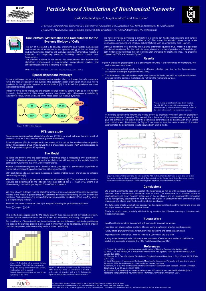
Figure 1. PTS system diagram. Figure 4. Simple membrane-bound decay reaction. (Am +B C) Notice the different decay rate at the membrane (x=1), and the horizontal shift as a result of the errors introduced by the membrane reactions. Particle-based Simulation of Biochemical Networks Jordi Vidal Rodríguez1, Jaap Kaandorp1 and Joke Blom2 1) Section Computational Science (SCS), University of Amsterdam(UvA), Kruislaan 403, 1098 SJ Amsterdam, The Netherlands 2)Center for Mathematics and Computer Science (CWI), Kruislaan 413, 1098 SJ Amsterdam, The Netherlands SiC CellMath: Mathematics and Computation for the Systems Biology of Cells The aim of the project is to develop, implement, and validate mathematical and computational techniques for the systems biology of the cell. Biologists and mathematicians together will formulate realistic mathematical models of metabolic and regulatory networks including intrinsic spatial non-homogeneity. The planned outcome of the project are computational and mathematical algorithms, implemented in auto-adaptive computational models, and simulation results for the functioning of living cells. URL: www.siliconcell.net/sica/NWO-CLS/CellMath We have previously developed a simulation tool which can handle bulk reactions and surface (membrane) reactions as well as diffusion [4]. The space discretisation allows us to tackle inhomogenous mediums and introduce cellular features such as a membrane (see Figure 2). Blom [2] studied the PTS pathway with a partial differential equation (PDE) model in a spherical domain and membrane. For this particular case where the number of particles is sufficiently large, both models should produce the same results after averaging the stochastic ones. The gradients obtained by PDE’s are shown in Figure 3. • Results • Figure 4 shows the gradient profile of a decay reaction where A are confined to the membrane. We notice two sources of errors: • The membrane-cytosol reaction have a different effective rate due to the homogeneous assumption of Gillespie algorithm(vertical shift). • The diffusion of released membrane particles causes the horizontal shift as particles diffuse on average from the center of the lattice site, not from the membrane surface. Spatial-dependantPathways In many pathways part of its substances are transported along or through the cell’s membrane while the rest are located in the cytosol. This particular spatial organization might give rise to gradients in the cytosolic substances concentrations [2]. It is found that gradients are more significant for larger cells [5]. Moreover while some molecules are present in large number, others might be in low number increasing the fluctuations of the system. In some cases these might not be properly modelled by a system of PDEs, which are based on the mass action law reactive dynamics. In the more complex PTS network the results are not as expected: We do not observe gradients in the concentrations of proteins. We suspect this is because of the previous two sources of error plus the stiffness of the system and the geometrical error induced by discretisation of an sphere into cubical boxes. Neverthless, in Figure 5 we observe that the mass evolution of species approximates the deterministic results obtained with Blom’s model. PTScasestudy Posphoenolpyruvate:gyclose phosphotransferase (PTS) is a small pathway found in most of bacterias, such as E. coli, involved in the glucose metabolism. External glucose (Glc) is transported to the interior of the cell by the membrane-bound protein IICB.P. The phosporil group (P) is derived from a phsophoenolpyruvate (PEP) which is passed to the IICB protein through the PTS pathway. • The Model • To tackle the different time and space scales involved we chose a Mesoscopic level of simulation to avoid unaffordable molecular dynamics simulations yet still working at the particle level (in contrast to PDE models that work with concentrations). • Space is discretised regularly in a Cartesian lattice (see Figure 2). The diffusion of particles is carried out according Chopard multiparticle diffusion method [1]. • On each lattice site, an stochastic mesoscopic reaction method is run. Our choice is Gillespie reaction algorithm [2]. • Reaction and Diffusion processes are executed alternatively [6]. The duration of the reaction intervals are determined by the diffusion time step defined as: t = (1/2d) l2/D, where d is dimensionality, l is lattice spacing and D the diffusion coeficient. We have chosen Gillespie reaction algorithm because it is a computational feasible mesoscopic method of the Master Equation (ME) that describes the stochastic trajectory of a set of chemical reactions. First a reaction (m) is chosen following the probability distribution: Pr(m) = am/∑jaj,, where a is the propensity function. And then the virtual occurrence time (t) is assigned following the probability distribution: P(t) = ∑jaj exp(- t ∑jaj) dt. This method alone reproduces the ME results exactly, thus it can cope with any reaction system, provided it fulfils the requirements: reaction limited and well-stirred and initially homogeneous. For the diffusion, Chopard multiparticle method enhances the diffusion of particles by partitioning into groups the particles present a site i, and moving them to its neighbours, provided enough particles are present, otherwise each particle is moved individually. Figure 5. Mass evolution in time per species in the PTS system. Mass in shown on y axis, time on x axis. Stochastic results are shown in solid lines, while Deterministic are the dashed lines. Notice how some proteins decay or increase at different rate. These shifts might be originated on the membrane reactions. Conclusions We present a method to cope with spatial inhomogeneities as well as with stochastic fluctuations of reactions from a mesoscopic simulation point of view. The membrane is a principal source of inhomogeneity and error: Reaction processes on the membrane are only approximately simulated, due to homogeneity assumption on each lattice site implicit in Gillespie method, and diffusion also undergoes side effects from the fluxes through the membrane. The lattice size effects, which affects accuracy and computational cost, and the membrane errors are two major issues to research in the near future. Finally, in certain cases, specially with fast decay reaction, the diffusion time step t, interferes with the reaction process. • FutureWork • Modify (diffusion) method to cope with stiff systems by varying t parameter. • Combine non-planar surface and bulk diffusion using a cartessian grid, for membrane error. • Study lattice granularity effects for diffusion limited systems and complex geometries. • Hybridisation of the method: run best method at convenient site and time. • Using a membrane-cytosolic pathway where stochastic effects become evident to validate the spatial and stochastic properties that PDE models cannot account for. References 1) Chopard, B. and Droz, M. Cellular Automata Modeling of Physical Systems, Cambridge 1998. 2) Blom, J. G. and Peletier, M. A. Diffusive Gradients in the PTS System, Tech. report MAS-R00200, CWI Amsterdam 2000. 3) Gillespie, D. T. Exact Stochastic Simulation of Coupled Chemical Reactions, J. Phys. Chem. 81(25) 2340-2361, 1977. 4) Vidal Rodríguez, J. Mesoscopic Stochastic Modelling for Biochemical Networks with Membrane-bound Molecules, MSc thesis Universiteit van Amsterdam, September 2004. 5)Francke, C. et al. Why the Phosphotransferase System of Escherichia coli Escapes Diffusion Limitation. Biophysical J. vol 85, pp. 612-622, 2003. 6) Bormann, G. Aanpassing en implementatie van een MC methode voor reactie-diffusie in biofysisch realistiche compartimentele neuronmodellen, PhD thesis, Universiteit Antwerpen, 2001. Figure 2. Ilustration of a circular domain discretisation. Only 1/4th due to symmetry. Gray sites belong to membrane and cytosol, while yellow only to cytosol. Periodic boundary conditions are used due to symmetry of the circle. Figure 3. Gradients obtained with the deterministic PDE model by Blom (2). Membrane is located at x=1, center of spherical cell at x=0. Bottom-right figure are membrane-bound concentration in time. Project number NOW-CLS 635.100.007 as part of the Computational Life Sciences project at NWO. PhD students: Maciej Dobrzynski (CWI), Hanna Härdin (VU/CWI, per 1/4/05), Jordi Vidal Rodríguez (UvA) Staff: Joke Blom (CWI), Frank Bruggeman (VU), Jaap Kaandorp (UvA), Klaas Krab (VU), Mark Peletier (TU/e), Jan van Schuppen, (CWI/VU), Hans Westerhoff (VU)