Download

1 / 37
400 likes | 1.24k Vues
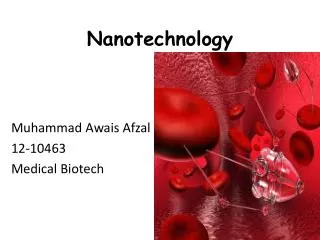
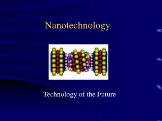
NANOTECHNOLOGY. Nanotechnology . WHAT IS NANOTECHNOLOGY ? According to National Nanotechnology Initiative (NNI) nanotechnology is defined as: "Nanotechnology is science,engineering,and technology conducted at nano scale or at molecular level ”

E N D
Nanotechnology • WHAT IS NANOTECHNOLOGY ? • According to National Nanotechnology Initiative (NNI)nanotechnology is defined as: "Nanotechnology is science,engineering,andtechnology conducted at nano scale or at molecular level” • nanotechnology is the study of extremely small things can be used across various fields. • Nano is a Greekword means “DRAWF” • Nanometer = 10-9= 1 billionth part of meter
HOW IT STARTED THERE’S IS A PLENTY OF ROOM AT THE BOTTOM "There's Plenty of Room at the Bottom" was a lecture given by physicist Richard Feynman at an American Physical Society meeting at Caltech on December 29, 1959.
Nanotechnology www.iub.edu.pk
NANOMEDICINE • DEFINITION:- • The simple definition of nano medicine is defined as the application of nanotechnology to the field of medicine . • But the definition according to the Medical Standing Committee of EUROPEAN SCIENCE FOUNDATION ESF • “The science and technology of diagnosing, treating, preventing disease and relieving pain of traumatic injury by preserving and improving human health, using molecular tools and molecular knowledge of the human body” .
Nano medicine and it’s branches NANOMEDICINE biology molecular medicine chemistry
CANCER TREATMENT Nano medicine
Why Nano medicine in cancer treatment • Target specific organelles within certain tissues or even the entire cells for localization in the targeted area. • Nanostructures can often overcome solubility and stability issues through surface modification/wrappings or additional formulation. • Nanostructures have novel physical properties, such as optical propertiesfor example quantum dots, which can be utilized for bio imaging. • high surface area high-dose therapeutic load can cause more devastating damage to cancer cells at the targeted site. • release therapeutic payloads by both active and passive transport to cancer sites significantly reduce nonspecific toxicity .
How nanoparticles reach the targeted Tumor cells • The nanoparticles reaches the targeted tumor cells by both active and passive transport. Active transport :- • nanoparticles that will actively target drugs to cancerous cells, based on the molecules that they express on their cell surface. • Molecules that binds to the particular cellular receptors on the surface of cancer cells will attach to a nanoparticle to actively target cells expressing the receptor. • Active targeting can even be used to bring drugs into the cancerous cell, by inducing the cell to absorb the Nano carrier. • Passive transport:- • Passive targeting occurs through a process known as "enhanced permeability and retention.(EPR)“. Because of the size and surface properties of nanoparticles can escape through blood vessel walls into tissues. • In addition, tumors tend to have leaky blood vessels and defective lymphatic drainage, causing nanoparticles to accumulate in them, thereby concentrating the attached cytotoxic drug where it's needed, protecting healthy tissue and greatly reducing adverse side effects. • Active targeting can be combined with passive targeting to further reduce the interaction of carried drugs with healthy tissue. Nanotechnology-enabled active and passive targeting can also increase the efficacy of a chemotherapeutic, achieving greater tumor reduction with lower doses of the drug.
Nano medicine in cancer treatment as Theranostics • Nanoparticles in Nanomedicine acts as both diagnostic and therapeutic agents • There are some nanoparticles which are most widely using as Nano carriers in Nanomedicine. • They are 1.liposomes 2.nanopores 3.quantumdots 4.fullerenes 5.dendrimers
Liposomes:- • Liposomes are tiny pouches made of lipid bilayeror fat molecules, surrounding by water core. • Liposomes are the first type of nanoparticles widely used for clinical cancer treatment. • During cancer treatment they encapsulate drugs, shield healthy cells from their toxicity, and prevent their concentration in vulnerable tissues such as those of a patient's kidneys and liver. • Liposomes can also reduce or eliminate certain common side effects of cancer treatment such as nausea and hair loss. • Examples:- • Doxorubicin HCl liposomal injection was the first liposomal encapsulated anticancer drug to receive clinical approval. • Lipoplatinis a new liposomal formulation of cisplatin, developed to reduce cisplatin toxicity, to improve drug accumulation at tumor sites and to overcome drug resistance. • Recently, DOX and fluoxetine coencapsulated liposomes have been reported to be effective formulation against drug-resistant MCF-7 cells. It was observed that liposomes significantly reduced tissue biodistribution of anticancer agents with improved cytotoxicity.
List of some liposomal formulations for cancer treatment (Table1=approved drugs, Table. 2 drugs in clinicaltrails)
Nanopores:- • Nanopores contains holes that are so tiny that DNA molecules can pass through them one strand at a time, allowing for highly precise and efficient DNA sequencing. • As a DNA strand moves through a nanopore, we can able to monitor each "letter" on it, deciphering coded information, including mutations associated with cancer. • By engineering nanopores into the surface of a drug capsule nanopores control the rate of a drug's diffusion in the body. • The size of Nano pores is 1.5 -20 nm • The size of DNA is approximately 2.5 nm because of this Nano size of DNA can easily pass through Nano pores which has a large size compare to DNA.
QUANTUMDOTS • Quantum dots are miniscule semiconductor particles. because they emit radiations at different wavelengths depending on the type of semiconducters used in their cores • cadmium sulfide for ultraviolet to blue • cadmium selenide for most of the visible • spectrumcadmium telluride for the far and near-infrared • A polymer coating enables attachment of molecules such as antibodies to tumors and other targeted cells. • The coating also shields nearby cells from the cadmium's toxicity. • The different colors of quantum dots provide a powerful tool for labeling and monitoring multiple cells and molecules simultaneously.
FULLERENS • These crystalline particles are a form of carbon atom whose molecular structure is arranged in a soccer ball-like structure. • They are also known as buckyballs. • They were discovered in 1985 among the detritus of laser-vaporized graphite. • They are used as cancer drug delivery vehicles, fullerenes don't break down in the body and are excreted intact. • This trait can be important for some cancer treatment compounds that are dangerous to healthy cells. • For example:- • Fullerene drug delivery particles that contain radioactive atoms would allow for the complete removal of radiation from the body following treatment.
Dendrimers • Dendrmimers are artificial man made molecule with potential to link treatment with detection and diagnosis. • The size of dendrimers is equall to the size of an average protein, and have a branching shape. • The branching shape gives them vast amounts of surface area to attach therapeutic agents or other biologically active molecules. • Researchers at the University of Michigan have fashioned dendrimers into sophisticated anti-cancer machines carrying five chemical tools • 1.Molecule designed to bind to cancer cells • 2.Emits fluoresces upon locating genetic mutation. • 3. Assist in imaging tumor shape using X-rays. • 4. Carries drugs released on demand. • 5. Sends a signal when cancerous cells are finally dead
Nanoparticles in molecularimaging The diagnostic aids of nanoparticles includes imaging of cancers 1. magnetic resonance imaging 2. Nuclear imaging 3. flouresence imaging 4. Ultrasound imaging
Magnetic resonance imaging Principle: MRI Step 1 :- Application of magnetic field by MRI scanner around area to be imaged Step2 :- exitation of hydrogen ions by using magnetic resonance energy causes emission of radiofrequency Step 3:- The emitted Radiofrequency produce an image based upon use of contrast agents which was read by machine Contrast agents are used to produce better image which are injected by intravenous route. 1.Comonly used contrast agent s are gadolinium (Gd)-based MRI agents due to it’s lanthanide’s ability
Role of nanoparticles in MRI • The contrast agents which are using in MRI are highly toxic and they don’t have any specificity towards target. • Due to acute toxicity and unspecificity of these agents leads to usage of nanprticles in MRI imaging. • example: • 1.Gadolinium-loaded single-walled carbon nanotubes are superparamagnetic, 40 timesmore efficient than traditional agents. • 2. Iron oxide nanoparticles (hundreds of nanometers) are super paramagnetic, while the Ultrasmall iron oxide nanoparticles (less than 50 nanometers) are more efficient.
Nuclear imaging POSITRON EMISSION TOMOGRAPHY • Uses positron emmitingnuclides such F18,11c and 64 cu • Emits high energy and posses high resonance capacity • Limited halflife radionuclides • SINGLE PHOTON EMMISION COMPUTED TOMOGRAPHY • Uses gamma rays emitting radio nuclides such as In 111, I 123 and m99 Tc • Emits low energy and posses low resonance capacity • long life radionuclides and high t1/2
Examples of how the nanoparticles used in neuro imaging 1.Fluorodeoxyglucose (18FDG) is the most commonly used radionuclide in PET imaging . As the analog of glucose, FDG can be used to detect cancer, which has a high glucose metabolic rate . FDG is not always sensitive to all lesions.Due to the potential toxicity of radiopharmaceuticals,nanoparticulate systems have been used to transfer in concentrated amounts 2. 11C-acetateis more suitable for the detection of liver cancer
Flourosence imaging PRINCIPLE • The measurement of flouresence producded by the molecules in near infrared region due to the excitement at specific wavelenght. • The NIR measures at wavelenght of 650-900 nm. • Advantages:- • 1. Sensitivity • 2. Multicolor detection • 3. Stability • 4. Low hazard • 5. Low cost Disadvantages:- • poor signal due to tissue autofluorescence examples: The combination of nanoparticle and fluorescent probe greatly enhances imaging . Quantum dot, which is referred to as semiconductor nanocrystals, can be used in fluorescence image, even to image the vasculature near tumor probe.
Ultrasound imaging Principle • Ultrasound travels freely through fluid and soft tissues. However, ultrasound is reflected back (it bounces back as 'echoes') when it hits a more solid (dense) surface. • So, as ultrasound 'hits' different structures of different density in the body, it sends back echoes of varying strength. probe can be used to calculate depth and help create a computer interpreted image of the scanned material. Because of physical differences in tissues density, features such as blood flow, organs, and tumors may be distinguished by ultrasound imaging. Example:- To visualize microvascular, nano/ microcapsules have been designed as a contrast agent for ultrasonic imaging for example Perfluorocarbons
Disadvantages of nanoparticles involvement in cancer therapy • The disadvantages of nanoparticle targeted delivery is very limited however there are a few disadvantages. They are • 1. The vasculature of tumor cells leads to extravasion and leakage of lymph vessels produce an heterogeneous blood flow. • 2. Due to lack of proper lymphatic drainage, the pressure of interstitial fluid increases continuously which may lead to hypertension of interstitial fluid. • 3. The increased intestinal fluid alter the transport capacity of extracellular surface by suppressing its diffusion tendency.
Some applications of nanoparticles in cancer treatment • Theranostic imaging of 5-fluorocytosine(5-FC) in prostate cancer by using siRNA. • For theranostic imaging of prostate cancer (PCa) ,a therapeutic nanoplex containing multimodal imaging was devoloped to target prostate-specific membrane antigen (PSMA), which is expressed on the cell surface of castrate-resistant PCa. • The nanoplex was designed to deliver small interfering RNA (siRNA) along with a prodrug enzyme to PSMA-expressing tumors. • Studies performed using two variants of human PC3-flu cells and PC3-pipcells, one with negative PSMA expression level and another with positive expression levels, demonstrated PSMA-specific uptake. • In addition, down-regulation of the selected siRNA target, choline kinase (Chk), and the conversion of the nontoxic 5-fluorocytosine(5-FC) to 5-flourouracil were also demonstrated with noninvasive imaging. • The nanoplex was well-tolerated and did not induce liver or kidney toxicity or a significant immune response.
Effect of supermagneticironoxide(SPIO) and anticancer drug paclitaxel in cancer imaging Paclitaxel (PTX)/super paramagnetic iron oxide (SPIO)-loaded PLGA-based nanoparticles for a theranostic purpose • SPIO did not show any toxicity in CT-26 cells while PTX-loaded nanoparticles had a cytotoxic activity on CT-26 cells. • PTX-loaded nanoparticle (5 mg/kg) with or without co-encapsulated SPIO induced in vivo shown a regrowth delay of CT26 tumors. • Significant cellular uptake by CT-26 cells of nanoparticles was shown by Prussian blue staining and fluorescent microscopy. • Application of multifunctional nanoparticles such as SPIO(contrast agent) and PTX in Nano medicine (PLGA) will provide satisfactory results in molecular imaging, drug delivery and real-time monitoring of therapeutic response.
REFERENCES:- • Nanotechnology and definition’s from Publications of Institute of nanotechnology(NANOIT) and National nanotechnology initiative(NNI) • Definition of Nano medicine-international journal of Nano medicine • Image of Nano medicine – www.vomts.arc.ulca.edu • Type of fields in Nano medicine and Nano medicine role in cancer – journal of Nano medicine by handwai • Active and passive transpor- http://ts1.eee.hku.hk/ccst9015sp13/p13/drug-delivery/cantcer-3/ nanotechnology in medicine. • Types of Nano carriers- http://www.pbs.org/wgbh/nova/sciencenow/3209/03-canc-nf.html link of Nano carrier Department of Bioorganic Chemistry, Faculty of Chemistry, Wrocław University of Technology, Wroclaw, Poland http://www.chemikinternational.com/year-2013/year-2013-issue-2/nanocarriers-in-medicine-siemieniec-j. • nanophore images and details www.pitys.org & national cancer institutes understanding cancer series. • MRI – journal of Nano medicine Hindwai
9. Nuclear imaging techniques – Neuroimaging perception lecture by David Heeger 10. Ultrasound imaging - patient.co.uk ultrasound scan. 11. Disadvantages of nanoparticles – from Class and slides of professor BRACCI 12. Theranostic imaging of 5-fluorocytosine(5-FC) in prostate cancer by using siRNA. 2012 Sep 25;6(9):7752-62. Epub 2012 Aug 9 and JHU ICMIC Program, Division of Cancer Imaging Research, The Russell H Morgan Department of Radiology and Radiological Science, The Johns Hopkins University School of Medicine, Baltimore, Maryland 21205, United States. 13. Paclitaxel (PTX)/super paramagnetic iron oxide (SPIO)-loaded PLGA- based nanoparticles for a theranostic purpose by 2013 Apr 15;447(1-2):94-101. doi: 10.1016/j.ijpharm.2013.02.042. Epub 2013 Feb 26. And Université Catholique de Louvain, Louvain Drug Research Institute, Pharmaceutics and Drug Delivery, Avenue Mounier, B1 73.12, 1200 Brussels, Belgium/ second application