Download
1 / 34
340 likes | 501 Vues
How E. Coli find their middle. Xianfeng Song Sima Setayeshgar. Outline. Introduction to E. coli Two systems regulate division site placement Nucleoid occlusion Min proteins Experiments: Qualitatively understand Min Proteins in E. coli Model: How and why Min Proteins work in E. coli ?.

E N D
How E. Coli find their middle Xianfeng Song Sima Setayeshgar
Outline • Introduction to E. coli • Two systems regulate division site placement • Nucleoid occlusion • Min proteins • Experiments: Qualitatively understand Min Proteins in E. coli • Model: How and why Min Proteins work in E. coli?
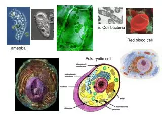
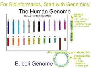
About E. coli • E. coli is a bacterium commonly found in the intestinal tracts of most vertebrates. • Studied intensively by geneticists because of its small genome size, normal lack of pathogenicity, and ease of growth in the laboratory. • Size: about 0.5 microns in diameter and 1.5 microns long From: cwx.prenhall.com
E. coli life cycle Typical lifecycle
E. coli cell division • Division accuracy: .50 +/- .02 • Placement of FtsZ ring: .50 +/- .01
Two systems regulate division site placement • Nucleoid occlusion • Min proteins • Including MinC, MinD, and MinE
Min proteins (Experiments) • MinC Inhibits FtsZ ring formation • MinD MinD:ATP recruits MinC to membrane • MinE Binds to MinD:ATP in membrane and induces ATP hydrolysis Black: MinC Red: MinD Blue: MinE
Without Min proteins, get minicelling phenotype (Min-) If MinC is over-expressed, get filamentous growth (Sep-) Min proteins (Experiments)
Min proteins oscillate from pole to pole Hale et al.(2001) MinD-GFP
MinE ring caps MinD polar region • MinE ring is membrane bound. • Ring appears near cell center and moves toward pole. Hale et al.(2001) MinE-GFP
Filamentous cell has “zebra stripe” pattern of oscillations • Wavelength of oscillations is ~10 microns. Raskin and de Boer(1999) Hale et al.(2001) MinD-GFP MinE-GFP
Phenomenology of Min oscillations • MinD polar regions grow as end caps. • MinE ring caps MinD polar region. • Oscillation frequency: • [MinE] frequency , • [MinD] frequency . • Filamentous cell has “zebra stripe” pattern of oscillations. • In vivo oscillations require MinD and MinE but not MinC.
How and Why Min-proteins oscillate (Modeling effort) • Howard et al. (2001) • Very simple 1D model • MinE is recruited by cytoplasmic MinD to membrane • MinD polar region fails to reform at poles. (not agree with experiment) • Huang and Wingreen (2003) • MinE is recruited by membrane-bound MinD-ATP • MinD aggregation on the membrane follow a one-step process • Kruse et al. (2005) • Consider protein diffusion on the membrane. • MinD aggregation on the membrane follow a two-step process: first attach to membrane, then self-assembles into filament • Meinhardt and de Boer (2001) • Requires new protein synthesis.
Result: MinD/E movie MinE MinD
Mechanism for growth of MinD polar regions (Huang and Wingreen, 2003) • MinD ejected from old end cap diffuses in cytoplasm. • Slow nucleotide exchange implies uniform reappearance of MinD:ATP. • Capture of MinD:ATP by old end cap leads to maximum of cytoplasmic MinD:ATP at opposite pole.
Result: Frequency of oscillations ~ [MinE]/[MinD] • No oscillations for [MinE] too high, or for [MinD] too low. • Minimum oscillation period 25s. 4 micron cell
Result: “Zebra stripe” oscillations in long cells • Stripes form with wavelength of ~10 microns
Oscillation makes E. Coli divide accurately • The oscillation make MinD has smaller average concentration at the middle and higher concentration at the ends. • MinC can only be bounded to memberane if it is recruited by MinD-ATP • MinC can Inhibits FtsZ ring formation, thus inhibits the division
Linear stability analysis around homogeneous solution Red curve corresponds to a normal cell
Predictions of model (Huang and Wingreen, 2003) • Delay in MinD:ATP recovery is essential. (Verified by Joe Lutkenhaus – nucleotide exchange rate is slow, ~2 / s.) • Rate of hydrolysis of MinD:ATP by MinE sets oscillation frequency. • Diffusion length of MinD before rebinding to membrane sets oscillation wavelength.
Recent development • Helical morphology of MinD polymers. • Some recent modeling effort (mainly focus on cleaning some details, no breakthrough) • the fluctuation effects on the min proteins (Howard, et al, 2003), • The new model considering the membrane diffusion and more reactions (Kruse, et al, 2005) • min-protein oscillations in round bacteria (Huang and Wingreen, 2004)
Open questions • MinE ring moving in reverse. • The helical structure of MinD polymers
Conclusions • Although bacteria such as E. coli is a very simple creature, it is also very complicated system. • To understand this system, physicist can help a lot.
Evidence from in vitro studies A. Phospholipid vesicles B. MinD:ATP binds to vesicles and deforms them into tubes • MinD:ATP polymerizes on vesicles • Diffraction pattern indicates well-ordered lattice of MinD:ATP E. MinE induces hydrolysis of MinD:ATP and disassembly of tubes Hu et al. (2002)
Min proteins in spherical cells:Neisseria gonorrhoeae Szeto et al. (2001) Wild type MinDNg-
Why does E. coli need an oscillator? In B. subtilis, minicelling is prevented by MinCD homologs, but polar regions are static. Marston et al. (1998)
How E. coli find its middle Proteins are too small to see the caps’ curvature Subtilis have local proteins fixed at two ends, but E. coli does not have