Download
1 / 28
760 likes | 2.33k Vues
Venous Leg Ulcers. Why do patients with chronic venous insufficiency develop VLU?. CVI most common cause of VLU VLU patients have venous hypertension, or abnormally sustained elevation of venous pressure on walking Caused by vein valve reflux, outflow problems or both

E N D
Why do patients with chronic venous insufficiency develop VLU? • CVI most common cause of VLU • VLU patients have venous hypertension, or abnormally sustained elevation of venous pressure on walking • Caused by vein valve reflux, outflow problems or both • Venous outflow issues • Venous obstruction • Poor function of calf muscle pump impairs venous system's ability to return venous blood to heart • Ankle movement limitations contribute to calf muscle pump failure
What are the risk factors for VLU? • Age older than 55 years • Family history of CVI • Ulcer history, parental history of ankle ulcers • Higher body mass index • History of pulmonary embolism • Venous reflux in deep veins, history of superficial/DVT • Lower extremities skeletal or joint disease • Number of pregnancies • Physical inactivity • Severe lipodermatosclerosis
Are there measures that can prevent VLU or their recurrence? • Aggressive management of reversible risk factors • Control of relevant comorbid conditions (CHF, PVD) • Healthy diet, appropriate exercise, weight control • Management of a hypercoagulable state • Stockings that achieve at least 20-30 mm Hg pressure • Patients should use highest level of compression tolerable • Surgical venous ablation
CLINICAL BOTTOM LINE: Prevention... • CVI is the leading cause of VLU • Venous hypertension with calf muscle pump dysfunction • Manage comorbid risk factors • CVI, obesity, hypercoagulable states • Skeletal and joint disease of the lower extremities • Compression stockings • For primary and secondary prevention • Venous intervention • For secondary prevention
What symptoms and physical findings are suggestive of CVI? • Swelling and aching of legs, worse at end of day and improved by leg elevation • History of ulcer recurrence, particularly at same location • Dependent edema, telangiectasias, varicose veins, reddish-brown pigmentation and purpura, and subsequent hemosiderin deposition • Eczematous changes with redness, scaling, pruritus • Smooth, ivory-white, stellate atrophic plaques of sclerosis with telangiectases (atrophie blanche) • Chronic lipodermatosclerosis (LDS) and acute LDS
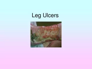
What symptoms and physical findings suggest that VLU are due to CVI? • VLU may be painful—dull, aching, or burning pain • Location over medial lower third of the legs • Usually 1 ulcer w/ irregular, flat, or only slightly steep borders • Ulcer bed shallow, with granulation tissue or fibrinous material • Wound surface rarely shows necrosis, exposed tendons, bone • Venous dermatitis, LDS, or atrophie blanche around ankle • Assessment: Test for neuropathy • Severity of CVI correlates with decreased range of motion at ankle and is associated with peripheral neuropathy • VLU pain neuropathic in origin in some patient
What other conditions should be considered during evaluation of a patient with possible VLU? • Common causes of lower extremity ulcers • CVI • Arterial insufficiency • Diabetic neuropathy • Prolonged pressure • Less common causes • Trauma • Inflammatory or metabolic conditions • Cancer • Infections
What is the role of laboratory testing? • No single laboratory test is diagnostic • Testing may be indicated depending on specific patient history, comorbidities, and family history • In patients with history of recurrent ulceration or thrombosis, evaluate for hypercoagulable states
What is the role of noninvasive tests, such as ankle-brachial index and duplex ultrasonography? • Ankle-brachial index should be performed • For PAD screening: concomitant arterial disease in ~20% • Compression therapy could worsen an arterial ulcer • Color duplex ultrasonography • For accurate diagnosis and to provide prognostic info • Photo and air plethysmography • Whole-limb venous hemodynamics at rest and after exercise • CT exam • Intractable edema associated with pain despite compression
What is the role of routine testing for infection? • Swab culture testing unwarranted w/o signs of infection • If atypical infection suspected: send tissue from wound biopsy for microscopic examination and culture • Use antibiotic therapy only for clinically infected ulcers • Evidence supports topical cadexomer iodine for healing • No evidence supports use of systemic antibiotics
When should clinicians consider obtaining a biopsy or referring the patient to a surgical or nonsurgical specialist for diagnosis? • To rule out other causes of VLU, especially cancer • When ulcers are atypical-appearing ulcers • When ulcers have not healed after 4 weeks of active treatment
CLINICAL BOTTOM LINE: Diagnosis... • Typically based on clinical history and physical examination • Presence of CVI • Single, painful ulcer with irregular, flat borders and granulating or fibrinous bed on medial lower third of legs • Color duplex ultrasonography to characterize venous disease in all patients • Ankle-brachial index to exclude concurrent PAD • If VLU do not improve within 4 weeks of active therapy: consider referral to specialist or biopsy
What is the overall approach to treatment? • Treatment goals • Reduce edema and pain • Heal ulcers • Prevent recurrence • Systematic approach needed • Assess frequently and escalate treatment if unresponsive • Simplest treatment: bed rest with leg elevation • Elevate legs above heart 30 minutes, 3 to 4x/d + at night • Reduces swelling, improves venous microcirculation • Most patients struggle to follow this recommendation
What is the role of compression therapy? • Cornerstone of therapy • Because sustained leg elevation often difficult to achieve • Gold standard: multiple elastic layers for graduated compression • Increases interstitial hydrostatic pressure • Improves venous return • Reduces venous hypertension and edema • Improves ulcer healing rates • Use cautiously with CHF and with arterial insufficiency • Don’t use with severe arterial insufficiency
How long should clinicians prescribe compression therapy? • Continue until the ulcer heals • Continue indefinitely after healing to prevent recurrence • To enhance adherence, instruct how to put on stockings • Ensure proper measurement and fit • Assistive devices may help arthritic, obese, elderly patients • Replace at least every 6 months
What is the role of medication? • To improve healing in combination with compression • Aspirin (300 mg daily) • Pentoxifylline (400-800 mg 3x/d) • To reduce LDS inflammation, pain, induration • Stanozolol • Oxandrolone • Horse chestnut seed extract (active ingredient: aescin) • To reduce pain (based on neuropathic origin) • Amitriptyline, gabapentin, pregabalin
What is the role of growth factors? • Granulocyte macrophage colony-stimulating factor • Topical and perilesional injection increases ulcer healing • Promotes wound healing through many mechanisms (homeostasis, inflammation, proliferation, maturation) • Increases vascularization • FDA-approved for neutropenia but not wound healing • Phase 3 trials stopped due to bone pain associated with perilesional injections
What is the role of physical therapy or exercise? • Aim: to improve range of ankle movement and calf muscle pump function • Might enhance ulcer healing • But evidence conflicting and RCTs lacking • RCT underway: comparing compression therapy with compression therapy + 12 weeks of supervised exercise
What is the role of hyperbaric oxygen therapy? • Adjunct to standard wound care • Controversial because evidence for treating VLU extremely limited • 100% oxygen at 2-2.5 atmosphere absolute for 60- to 120-minute periods over 15-30 sessions • Goal: increase partial pressure of oxygen at the wound • Role in pathogenesis and treatment unclear • Fibrin cuff theory: fibrin cuffs formed around precapillary vessels may result in wound hypoxia, so increased oxygen might aid healing
What is the role of surgical debridement or skin grafting? • Debridement • Removes nonviable tissue to achieve an appropriate wound bed with granulation tissue • Standard care despite lack of controlled data on healing • Skin grafting • Enhances healing for large or slow-healing ulcers • May rapidly decrease pain and aid functional status • Pinch grafts, split-thickness skin grafts, and micro-skin grafts used successfully but RCTs lacking • Skin equivalents (cellular, acellular) may aid healing
What is the role of venous surgery in treatment and prevention? • Venous surgery • Doesn’t improve healing but reduces recurrence • Open surgery has significant potential morbidity • Cochrane review found no evidence for benefit or harm • Subfascial endoscopic perforator surgery • Safer, possible improved healing, decreased recurrence • Minimally invasive procedures • Treat CVI and recurrence • Endovenous thermal ablation (laser, radiofrequency, steam) • US-guided foam sclerotherapy; cyanoacrylate embolization
When should clinicians consider referring the patient to a surgical or nonsurgical specialist for treatment? • Prognostic factors associated with slower healing • Larger wound area (>5 cm2) and long duration (>6 months) • LDS and ulcer history, BMI >33 kg/m, physical inactivity • Prolonged venous filling time, deep venous insufficiency • Ulcer depth >2 cm, atypical ulcer location (posterior calf) • Refer to wound specialist when wounds fail to decrease in size during first month of treatment • Expertise may be found in a variety of specialties • Vascular medicine and surgery, podiatry, dermatology
How should clinicians educate patients? • Encourage patients to adhere to compression therapy • Provide educational materials on pathophysiology, management, and prevention • Consider video-based educational interventions to teach patients about the disease • Consider patient support groups for education on self-management
CLINICAL BOTTOM LINE: Treatment... • Goals: reduce edema, improve pain and LDS, heal ulcer, prevent recurrence • Maintenance: • Moist wound bed and regular sharp debridement • Infection control • Compression with elastic multilayer bandages • If no improvement in 4 weeks: consider referral to wound expert and adjuvant therapies • Prevent recurrence: indefinite use of compression stockings and vascular intervention