Download
1 / 31
310 likes | 335 Vues
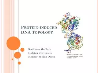
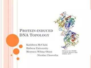
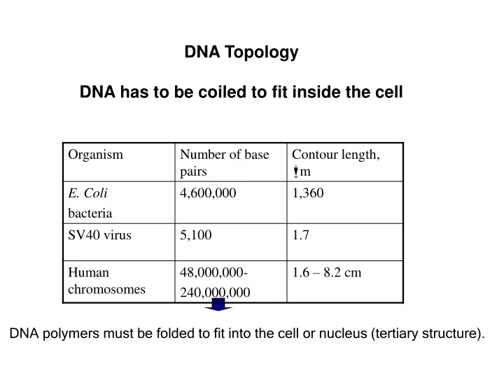
DNA Topology DNA has to be coiled to fit inside the cell. DNA polymers must be folded to fit into the cell or nucleus (tertiary structure). DNA Topology :. DNA Topology: linking number. Consider a 260 bp B-duplex:. Connect the ends to make a circular DNA:. Tw = 260/10.4 = 25.

E N D
DNA Topology DNA has to be coiled to fit inside the cell DNA polymers must be folded to fit into the cell or nucleus (tertiary structure).
Connect the ends to make a circular DNA: Tw = 260/10.4 = 25
An electron micrograph of negatively supercoiled and relaxed DNA Stryer Fig. 27.20
Organization of chromosomal DNA • Chromosomal DNA is organized in loops (no free ends) • It is negatively supercoiled: 1 (-) supercoil per 200 nucleotides 145 bp duplex Histone octamer (H2A, H2B, H3, H4)2 H1 is bound to the linker region
Enzymes that control DNA supercoiling: DNA Topoisomerases Change the linking number (Lk) of DNA duplex by concerted breakage and re-joining DNA strands Topoisomerase enzymes Topoisomerases I Relax DNA supercoiling by increments of 1 (cleave one strand) Topoisomerases II Change DNA supercoiling by the increments of 2 (break both strands) Usually introduce negative supercoiling
Human DNA Topoisomerase I: DNA: side view 20Å Stryer Fig. 27.21
723 Mechanism of DNA Topoisomerases I OH P-Topo Wr = 1
Drugs that inhibit DNA Topoisomerase I • • Camptothecin, topotecan and analogs • •Antitumor activity correlates with interference with topoisomerase activity • • Stabilizes topoisomerase I-DNA intermediate, preventing DNA strand re-ligation • Used in treatment of colorectal, ovarian, and small cell lung tumors
Enzymes that control DNA supercoiling: DNA Topoisomerases Change the linking number (Lk) of DNA duplex by concerted breakage and re-joining DNA strands Topoisomerase enzymes Topoisomerases I Relax DNA supercoiling by increments of 1 (cleave one strand) Topoisomerases II Change DNA supercoiling by the increments of 2 (break both strands) Usually introduce negative supercoiling
Topoisomerases II • Most of Topoisomerases II introduce negative supercoils (e.g. E. coli DNA Gyrase) • Require energy (ATP) • Each round introduces two supercoils ( Wr = - 2) • Necessary for DNA synthesis • Form a covalent DNA-protein complex similar to Topoisomerases I
Yeast DNA Topoisomerase II Stryer Fig. 27.23
Topoisomerase II - mechanism Stryer Fig. 27.24
Drugs that inhibit bacterial Topoisomerase II (DNA gyrase) Interfere with breakage and rejoining DNA ends: Inhibit ATP binding:
Enzymes that cut DNA and RNA : nucleases (Dnases and Rnase) Phosphate group Nucleobase 2’-deoxyribose HO Exonucleases A 5’ 3’ 5’ 3’ 5’ + dNMPs • Degrade DNA in a stepwise manner by removing deoxynucleotides in 5’ 3’ (A) or 3’ 5’ direction (B) • Require a free OH • Exonucleases can be active on both single- and double-stranded DNA • Used for degrading foreign DNA and in proofreading during DNA synthesis B 3’ Examples: B: snake venom phosphodiesterase A : calf intestinal phosphodiesterase
DNA Endonucleases • Cleave internal phosphodiester bonds resulting in 3’-OH and 5’-phosphate ends 5’ 3’-OH 5’-P 5’-P 3’-OH • some endonucleases cleave randomly (DNase I, II) • Type II Restriction endonucleases are highly sequence specific EcoRI recognition site: Palindromic site (inverted repeat) • RE are found in bacteria where they are used for protection against foreign DNA
Recognition sequences of some common restriction endonucleases
DNARestrictionEnzyme EcoR V H N O N NH 2 O N N HN N N O N O A•T 5’-GAT ATC-3’ 3’-CTA TAG-5’ Asn185 Thr186
Applications of Restriction Endonucleases in Molecular Biology • DNA fingerprinting (restriction fragment length polymorphism). • 2. Molecular cloning (isolation and amplification of genes).
Restriction fragment length polymorphisms are used to compare DNA from different sources
DNA Ligase O -O P O DNA Ligase + O- (ATP or NAD+) AMP + PPi O O P O OH O- • Forms phosphodiester bonds between 3’ OH and 5’ phosphate • Requires double-stranded DNA • Activates 5’phosphate to nucleophilic attack by transesterification with activated AMP
Human Genetic Polymorphisms • Human genome size: 3.2 x 10⁹ base pairs • 30,000 genes • 2-4 % of total sequence codes for proteins • Human genetic variation: • 1 sigle nucleotide polymorphism (SNP) per 1,300 bp
Examples of genetic polymorphisms of drug metabolizing enzymes Enzyme substrate examples DNA regions involved cytochrome 2B6 cyclophosphamide exons 1,4,5, and 9 tamoxifen benzodiazepines cytochrome 2D6 debrisoquine internal base changes cytochrome 1A2 caffein 5' flanking region phenacetin N-acetyltransferase aromatic amines
DNA Structure: Take Home Message • Genetic information is stored in DNA. • DNA is a double stranded biopolymer containing repeating units of nitrogen base, deoxyribose sugar, and phosphate. • DNA can be arranged in 3 types of duplexes which contain major and minor grooves. • DNA can adopt several topological forms. • There are enzymes that will cut DNA, ligate DNA, and change the topology of DNA. • Human genome contains about 3.2 billion base pairs. Inter-individual differences are observed at about 1 per 1,000 nucleotides.