Download
1 / 61
620 likes | 788 Vues
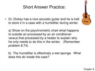
Answer to Practice Problem Draw the chemical structure of the tripeptide Ala – Ser – Cys at pH 7. Answer the following with regard to this tripeptide: 1. Indicate the charge present on any ionizable group(s). 2. Indicate, using an arrow, which covalent bond is the peptide bond.

E N D
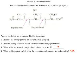
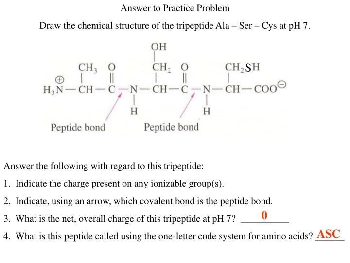
Answer to Practice Problem Draw the chemical structure of the tripeptide Ala – Ser – Cys at pH 7. Answer the following with regard to this tripeptide: 1. Indicate the charge present on any ionizable group(s). 2. Indicate, using an arrow, which covalent bond is the peptide bond. 3. What is the net, overall charge of this tripeptide at pH 7? __________ 4. What is this peptide called using the one-letter code system for amino acids? ______ S 0 ASC
Proteins: Three Dimensional Structure and Function
Ribbon diagram Space-filling model Figure 4.3
Levels of Protein Structure Figure 4.1
Resonance structure of the peptide bond Figure 4.5
Planar peptide groups in a polypeptide chain Figure 4.6
Trans and cis conformations of a peptide group Figure 4.7 Nearly all peptide groups in proteins are in the trans conformation
Rotation in a peptide Figure 4.8 phi psi N-Ca Ca-C
Ramachandran Plot Figure 4.9
The alpha helix Figure 4.10
The alpha helix Figure 4.11
An amphipathic alpha helix Figure 4.12
Amphipathic alpha helices are oftenfound on the surface of a protein Figure 4.13
The beta sheet Parallel Figure 4.16
The beta sheet Parallel Figure 4.16 N N N
The beta sheet Antiparallel Figure 4.16
The beta sheet Antiparallel Figure 4.16 N N N
The beta sheet. Side chains alternatefrom one side to another Figure 4.17
Levels of Protein Structure Figure 4.1
Reverse turns Figure 4.19 Type II b turn Type I b turn
Reverse turns Figure 4.19 Type II b turn Type I b turn
Supersecondarystructures,often called“motifs” Figure 4.20
Domain foldsin proteins Figure 4.25
QuaternaryStructure Figure 4.26
How do proteins fold and unfold? The information for proteins to fold is contained in the amino acid sequence. Can proteins fold by themselvesor do they need help?
Intermediates inprotein folding Figure 4.37
Heating proteins willunfold or “denature”the molecule. Figure 4.31
Anfinsen’sprotein foldingexperiment Figure 4.35
Protein Folding A cell can make a biologically active protein of 100 amino acids in 5 seconds. If each amino acid could adopt 10 different conformations this makes 10100 different conformations for the protein. If each conformation were randomly sampled in 10-13 seconds it would take 1077 years Therefore protein folding must not be a random process.
Energy well of protein folding Figure 4.36
Forces driving protein folding: • Hydrophobic effect • Hydrogen bonding • Charge-charge interactions • Van der Waals interactions
Molecular Chaperones(Chaperonins) Some proteins don’t spontaneously fold to native structures. They receive help from proteins called chaperonins Best characterized chaperonin system is from E. coli. GroEL / GroES chaperonin system (GroE chaperonin) These chaperonins bind to unfolded or partially folded proteins and prevent them from aggregating. They assist in refolding the proteins before releasing them.
GroE Figure 4.38
Chaperonin-assisted protein folding Figure 4.39
Three-dimensional structuresof specific proteins 1. Collagen, a fibrous protein 2. Myoglobin and Hemoglobin, O2 binding proteins 3. Antibodies
Collagen is a fibrous proteinfound in vertebrateconnective tissue. Collagen has a triple helixstructure, giving it strengthgreater than a steel wire ofequal cross section.
Collagen is 35% Glycine 21% Proline + Hydroxyproline The repeating unit is Gly – X – Pro (HyPro) The interior of a collagen triple helix is packed with Glycines (red)
4-Hydroxyproline and 5-Hydroxylysine residues Figure 4.41 and 4.43
Allysine and lysine residues form cross-links in collagen Figure 4.44
Allysine residues form cross-links in collagen Figure 4.44
Hemoglobin and Myoglobin bind oxygen Figure 4.46 Heme Histidines Protein