Download
1 / 74
740 likes | 1.03k Vues
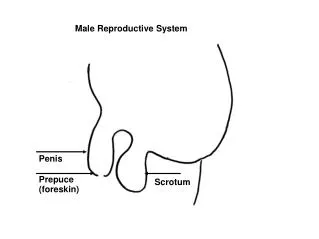
Male Reproductive System. The organs of the reproductive system are Internal reproductive organs External genital organs. Primary Sex Organs (Gonads). The primary reproductive organs or gonads consist of a pair of testes in the male and a pair of ovaries in the female.

E N D
The organs of the reproductive system are • Internal reproductive organs • External genital organs
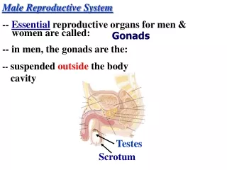
Primary Sex Organs (Gonads) • The primary reproductive organs or gonads consist of a pair of testes in the male and a pair of ovaries in the female. • In both sexes the mature gonads perform the dual function of • producing gametes (gametogenesis), that is, spermatozoa (sperm) in the male and ova (eggs) in the female • Secreting sex hormones testosterone in males and estrogen and progesterone in females. • In addition to the gonads the reproductive system in each sex includes a reproductive tract which is a system of ducts that are specialized to transport or house the gametes after they are produced plus accessory sex glands that empty their supportive secretions into these passageways
The major male accessory sex glands whose secretions provide the bulk of the semen are • Seminal vesicles • Prostate gland • Bulbourethral glands
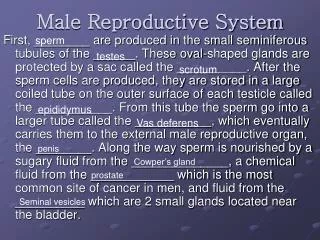
Sperm exit each testis through the male reproductive tract consisting on each side of an epididymis, ductus (vas) deferens, and ejaculatory duct • These pairs of reproductive tubes empty into a single urethra, the canal that runs the length of the penis and empties to the exterior
The secondary sexual characteristics are the external characteristics not directly involved in reproduction that distinguish males and females such as body configuration and hair distribution
Physiologic Anatomy • Testis is composed of up to 900 coiled seminiferous tubules, each averaging more than one-half meter long, in which the sperm are formed • The sperm then empty into the epididymis, another coiled tube about 6 meters long • The epididymis leads into the vas deferens, which enlarges into the ampulla of the vas deferens immediately before the vas enters the body of the prostate gland
Two seminal vesiclesone located on each side of the prostate empty into the prostatic end of the ampulla and the contents from both the ampulla and the seminal vesicles pass into an ejaculatory duct leading through the body of the prostate gland • Ejaculatory duct empty into the internal urethra.
The urethra is the last connecting link from the testis to the exterior • The urethra is supplied with mucus derived from a large number of minute urethral glands located along its entire extent and from bilateral bulbourethral glands (Cowper glands) located near the origin of the urethra
Sertoli Cells • Sertoli cells are large • They are present inside seminiferous tubule • The spermatogenic cells are attached to Sertoli cells by means of cytoplasmic connection • Sertoli cells support and nourish the spermatogenic cells • Secrete Mullerian regression factor • Secrete Inhibin and Activin • Secrete androgen binding protein and estrogen binding protein
Blood-Testis Barrier • It is a mechanical barrier that separates blood from seminiferous tubules of the testes • It is formed by tight junctions between the adjacent Sertoli cells near the basal lamina of seminiferous tubule • It protects the seminiferous tubules and spermatogenic cells by preventing the entry of toxic substances from blood into testis • It permits the nutritive and other essential substances to pass through
Rete Testis • Each seminiferous tubule opens into a network of thin walled channels called Rete Testis
Interstitial Cells of Leydig • These are the hormone secreting cells of the testis situated between the seminiferous tubules
Spermatogenesis • Spermatogenesis occurs in the seminiferous tubules during active sexual life as the result of stimulation by anterior pituitary gonadotropic hormones, beginning at an average age of 13 years and continuing throughout most of the remainder of life but decreasing markedly in old age
Spermatogenesis • During formation of the embryo, the primordial germ cells migrate into the testes and become immature germ cells called spermatogonia • The spermatogonia begin to undergo mitotic division beginning at puberty and continually proliferate and differentiate through definite stages of development to form sperm
In the first stage of spermatogenesis the spermatogonia migrate among Sertoli cells toward the central lumen of the seminiferous tubule • Spermatogoniathen become progressively modified and enlarged to form large primary spermatocytes • The primary spermatocytes undergo meiotic division to form two secondary spermatocytes.After few days, these too divide to form spermatids that are eventually modified to become spermatozoa (sperm) • The entire period of spermatogenesis, from spermatogonia to spermatozoa takes about 74 days
Formation of Sperm • When the spermatids are formed they still have the usual characteristics of epithelioid cells but soon they begin to differentiate and elongate into spermatozoa • Each spermatozoon is composed of a head and a tail • On the outside of the anterior two thirds of the head is a thick cap called the acrosome that is formed mainly from the Golgi apparatus. This contains a number of enzymes similar to those found in lysosomes of the typical cell • These enzymes play important role in allowing the sperm to enter the ovum and fertilize it
Hormonal Factors That Stimulate Spermatogenesis • Testosterone( growth and division of the testicular germinal cells, which is the first stage in forming sperm) • Luteinizing hormone stimulates the Leydig cells to secrete testosterone • Follicle-stimulating hormone stimulates the Sertolicells, without this stimulation the conversion of the spermatids to sperm (the process of spermiogenesis) will not occur
Estrogens formed from testosterone by the Sertoli cells when they are stimulated by follicle-stimulating hormone are also essential for spermiogenesis. • Growth hormone (as well as most of the other body hormones) is necessary for controlling background metabolic functions of the testes. Growth hormone specifically promotes early division of the spermatogonia
Other Factors Affecting Spermatogenesis • Increase in body temperature inhibits spermatogenesis ( internal temperature of scrotal sac is 2 degrees centigrade lower than body temperature) • (Regulation of temperature of scrotal testes is brought about by relaxation or contraction of scrotal musculature in summers and winters respectively) • Infections such as mumps cause degeneration of seminiferous tubules
The two testes of the human adult form up to 120 million sperms each day • A small quantity of these can be stored in the epididymis but most are stored in the vas deferens • The normal motile, fertile sperm are capable of flagellated movement through the fluid medium at velocity of 1 to 4 mm/min • Sperm can live for many weeks in the male genital ducts, once they are ejaculated in the semen, their maximal life span is only 24 to 48 hours at body temperature. At lowered temperatures semen can be stored for several weeks
Function of Seminal vesicles • Each seminal vesicle is a tortuous, loculated tube lined with a secretory epithelium that secretes a mucoid material containing an abundance of fructose, citric acid and other nutrient substances as well as large quantities of prostaglandins and fibrinogen (bulk 60% of the semen, nutrition) • During the process of emission and ejaculation each seminal vesicle empties its contents into the ejaculatory duct shortly after the vas deferens empties the sperm
Role of Prostaglandins • Prostaglandins help in fertilization in two ways (1) by reacting with the female cervical mucus to make it more receptive to sperm movement (2) by causing backward, reverse peristaltic contractions in the uterus and fallopian tubes to move the ejaculated sperm toward the ovaries
Function of the Prostate Gland • The prostate gland secretes a thin, milky fluid (30% of the semen) that contains calcium, citrate ion, phosphate ion, a clotting enzyme, and profibrinolysin • Prostatic fluid is alkaline in nature • Slightly alkaline prostatic fluid helps to neutralize the acidity of the other seminal fluids during ejaculation and thus enhances the motility and fertility of the sperm
Capacitation of spermatozoa is required for fertilization of the ovum • Spermatozoa when they leave the epididymis, their activity is held in check by multiple inhibitory factors secreted by the genital duct epithelia. • On coming in contact with the fluids of the female genital tract multiple changes occur that activate the sperm for the final processes of fertilization. These collective changes are called capacitation of the spermatozoa. This normally requires 1 to 10 hours
Capacitation • The uterine and fallopian tube fluids wash away the various inhibitory factors that suppress sperm activity in the male genital ducts • The spermatozoa in the fluid of the male genital ducts are continually exposed to many floating CHOLESTEROL vesicles from the seminiferous tubules • This cholesterol is continually added to the cellular membrane covering the sperm acrosome, toughening the membrane and preventing release of its enzymes. • The sperm deposited in the vagina swim away from the cholesterol vesicles upward into the uterine cavity, and they gradually lose much of their other excess cholesterol over the next few hours. • As a result the membrane at the head of the sperm (the acrosome) becomes weaker
The membrane of the sperm also becomes more permeable to calcium ions • The calcium ions also cause changes in the cellular membrane that cover the leading edge of the acrosome making it possible for the acrosome to release its enzymes rapidly and easily as the sperm penetrates the layers surrounding the ovum
Acrosome Reaction • When the sperm reaches the zonapellucida of the ovum, the anterior membrane of the sperm binds specifically with receptor proteins in the zonapellucida • The entire acrosome rapidly dissolves and all the acrosomal enzymes are released. Within minutes these enzymes open a pathway for passage of the sperm head through the zonapellucida to the inside of the ovum. Within another 30 minutes the cell membranes of the sperm head and of the oocyte fuse with each other to form a single cell
Why Does Only One Sperm Enter the Oocyte? • Within a few minutes after the first sperm penetrates the zonapellucida of the ovum, calcium ions diffuse inward through the oocyte membrane and cause multiple cortical granules to be released by exocytosis from the oocyte into the perivitelline space. These granules contain substances that permeate all portions of the zonapellucida and prevent binding of additional sperm and they even cause any sperm that have already begun to bind to fall off
Cryptoorchidism • Failure of a testis to descend from the abdomen into the scrotum at or near the time of birth of a fetus
Male sex hormones (androgens) secreted by the testes are • Testosterone(most abundant) • Dihydrotestosterone • Androstenedione
Testosterone that becomes fixed to the tissues is converted within the tissue cells to dihydrotestosteroneespecially in certain target organs such as the prostate gland in the adult and the external genitalia of the male fetus • Some actions of testosterone are dependent on this conversion whereas other actions are not
Chemistry of Androgens • All androgens are steroid compounds • Both in the testes and in the adrenals, the androgens can be synthesized either from cholesterol or directly from acetyl coenzymeA
Testosterone is sereted from the interstitial cells of Leydig which lie in the interstices between the seminiferous tubules and constitute about 20 percent of the mass of the adult testes • Leydigcells are almost nonexistent in the testes during childhood • These cells are numerous in the newborn male infant for the first few months of life and in the adult male after puberty • At both these times the testes secrete large quantities of testosterone
97% of the Testosterone is in the bound form with albumin and sex hormone binding globulin • In certain target tissues much of the Testosterone is converted to Dihydrotestosterone such as in prostate gland in the adult and external genitalia of the male fetus
Degradation and Excretion of Testosterone • The testosterone that does not become fixed to the tissues is rapidly converted by the liver into androsterone and dehydroepiandrosterone and simultaneously conjugated as either glucuronides or sulfates. These are excreted either into the gut by way of the liver bile or into the urine through the kidneys.
Production of Estrogen in the Male • The concentration of estrogens in the fluid of the seminiferous tubules is quite high and plays an important role in spermiogenesis. This estrogen is believed to be formed by the Sertoli cells by converting testosterone to estradiol • Much larger amounts of estrogens are formed from testosterone and androstanediol in other tissues (such as skin and adipocytes) especially the liver accounting for as much as 80 percent of the total male estrogen production
Testosterone is responsible for the distinguishing characteristics of the masculine body • During fetal life the testes are stimulated by chorionic gonadotropin from the placenta to produce moderate quantities of testosterone throughout the entire period of fetal development and for 10 or more weeks after birth • No testosterone is produced during childhood until about the ages of 10 to 13 years. • Testosterone production increases rapidly under the stimulus of anterior pituitary gonadotropic hormones at the onset of puberty and lasts throughout most of the remainder of life
Testosterone During Fetal Life • Testosterone is secreted first by the genital ridges and later by the fetal testes and is responsible for the development of the male reproductive organs
Effect of Testosterone to Cause Descent of the Testes • The testes descend into the scrotum during the last 2 to 3 months of gestation when the testes begin secreting reasonable quantities of testosterone
Puberty • Puberty is usually referred to as maturation of reproductive system and the development of secondary sexual characteristics • The average age for onset of puberty is between 10-13 years • First change at puberty is enlargement of testes and scrotum and scrotum becomes pigmented and rugose • This is followed by increase in the size of penis, appearance of pubic hair (pubarche), enlargement of accessory sex glands, appearnce of secondary sexual characteristics • Early morning erections and discharge of seminal fluid start taking place • An acceleration of linear growth and increase in body weight
Puberty (cont) • The hormones responsible for the start of puberty changes are the androgens (especially Testosterone) secreted by the testes • The adrenal cortex also starts contributing androgens (adenarche-increase in androgen production at the onset of puberty)
Effect of Testosterone on Development of Adult Primary and Secondary Sexual Characteristics • After puberty increasing amounts of testosterone secretion causes the enlargement male genital organs • Testosterone causes the secondary sexual characteristics of the male to develop, beginning at puberty and ending at maturity
Effect on the Distribution of Body Hair • Testosterone causes growth of hair • over the pubis (2) upward along the linea alba of the abdomen sometimes to the umbilicus and above (3) on the face (4)on the chest
Baldness • Testosterone decreases the growth of hair on the top of the head • Baldness is a result of two factors • a genetic background • superimposed on this genetic background large quantities of androgenic hormones