Download
1 / 148
1.53k likes | 1.78k Vues
NURS 2140 Fluid and Electrolytes Acid Base and IV Therapy. Teresa Champion, RN MSN Metropolitan Community College Winter 2012. BODY COMPOSITION AND FUNCTION. PRIMARY FLUID = Water 60% total body weight (1 L = 2.2 lbs) 2 – 2.5 L of water per day TWO PRIMARY FLUID COMPARTMENTS

E N D
NURS 2140 Fluid and Electrolytes Acid Base and IV Therapy Teresa Champion, RN MSN Metropolitan Community College Winter 2012
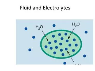
BODY COMPOSITION AND FUNCTION • PRIMARY FLUID = Water • 60% total body weight (1 L = 2.2 lbs) • 2 – 2.5 L of water per day • TWO PRIMARY FLUID COMPARTMENTS • Intracellular (ICF) • Extracellular (ECF) • FUNCTIONS: • Transporting • Removing • Regulation • Lubricating • Food Digestion
Regulation of Body Fluid • Osmosis – movement of water from lower particle concentration to higher particle concentration • Diffusion – movement of molecules from higher to lower concentration (simple or facilitated) • Filtration – movement of molecules through a semi-permeable membrane from higher concentration to lower concentration as a result of hydrostatic pressure
There are two ways in which substances can enter or leave a cell: • 1) Passive • a) Simple Diffusion • b) Facilitated Diffusion (carrier) • c) Osmosis (water only) • 2) Active • a) Molecules • b) Particles
Oncotic vs Hydrostatic Pressure • Filtration is directly opposed by the oncotic pressure of plasma proteins, especially Albumin in the blood stream. • Arteriole – high hydrostatic pressure - (~32mmHg) • Venus – low hydrostatic pressure – (~15 mmHg) • Plasma oncotic pressure – (~22mmHg) • http://www.youtube.com/watch?v=VMvD29-Agtg • http://www.youtube.com/watch?v=mpg7ON2CfFE • http://www.youtube.com/watch?v=dAO8igIysaA • Homeostasis - thus in a steady state ECF = ICF
OSMOLALITY • Number of molecules of solute per kg of water • NORMAL OSMOLALITY of blood is 275 – 295 milliosmoles per kg (mOsm/kg) of body weight • Isotonic Fluids - same osmolality as blood plasma) • Hypotonic Fluids - less concentration than blood plasma (< 275 mOsm/kg) • Hypertonic Fluids - greater concentration than blood plasma (>295 mOsm/kg)
Homeostatic Mechanisms • Fluid Balance - regulated by: • Osmoreceptors of the hypothalamus - stimulates release of ADH and stimulates thirst. • Baroreceptors (pressure sensitive cells) in carotids and aorta also stimulate the release of ADH • Baroreceptors in glomerular arterioles in kidney will secrete Renin and start the Renin-Angiotension (RAA) cascade thus resulting in release of aldosterone from the adrenal glands and cause sodium retention = fluid retention (water follows sodium)
Role of the Heart • Atrial Natriuretic Peptide: (ANP): secreted from atrial cells of heart (in response to too much volume in the blood) • acts as diuretic • inhibits thirst mechanism • suppresses the RAA cascade
Role of the Kidneys • Filter approx 180 Liters of blood per day; GFR (glomerular filtration rate) • Produces urine between 1-2 Liters/day • If loss of 1% to 2% of body water, will conserve water by reabsorbing more water from filtrate; urine will be more concentrated • If gain of excess body water, will excrete more water from filtrate; urine will be more diluted
Evaluation of Fluid Status • Normal serum hematocrit • 40 – 50% • Dilute serum • Low hematocrit and electrolyte levels • Concentrated serum • Elevated hematocrit and electrolyte levels
Nursing Considerations • Assess for headache, dizziness, syncope • CHF-SOB, dyspnea, activity intolerance • Maintaining accurate I & O • Daily weights • Monitoring Lab values • Frequency and consistency of stools • Meals include adequate fluid intake
Dehydration in the Elderly • Increased risks for dehydration: • Decrease in thirst • Lack of fluid replacement • Use of diuretic medications for high BP • Susceptibility to contagious diseases
Nursing Care Plan Dehydration Overhydration Risk for Imbalanced Fluid Volume Related to excessive fluid intake/decreased urination AEB …. • Risk for imbalanced Fluid Volume Related to excessive fluid loss/inability to take in fluids AEB ….
Electrolytes • Substance that develops an electrical charge when dissolved in water • Cation - positive charged • Anion - negative charged • Examples of cations: Sodium, Potassium, Magnesium, Calcium • Examples of anions: Chloride, Bicarbonate, Phosphorous
SodiumNormal serum values135-145 mEq/L • Most abundant cation in ECF • Functions • ECF volume – water balance • Acid-base balance • Nerve impulse control – sodium potassium pump • Levels below 115 mEq/L – brain damage, seizures • Sodium is primarily excreted through the kidneys, but other avenues are GI Secretions and sweat.
Sodium Deficit - Levels < 135 mEq/LHyponatremia • Loss through GI tract, skin or kidneys • An increased amount of sodium shift into the cells when there is a potassium deficit • An excessive ADH release (SIADH) causing Water retention and sodium deficit • Inadequate sodium intake, increased water intake • Excessive use of 5% dextrose solution • Levels below 115 mEq/L – brain damage
Pathophysiology of decreased sodium imbalances • CNS – excess water moves into the cerebral tissues – increased intracranial pressure • GI – loss causes acid base imbalances • Kidneys – renal dysfunction promotes sodium and water retention resulting in diluted sodium level (fluid overload) • Cellular activity - decrease in Na-K pump
Causes of Hyponatremia • Dietary changes – low sodium intake, excessive water intake, “fad diets”/fasting, anorexia nervosa, prolonged use of IV D5W • GI Losses – vomiting, diarrhea, GI Suctioning, Tap water enemas, GI surgery, Bulimia. • Renal Loses – Salt wasting kidney disease, diuretics. • Hormonal Influences - ADH, SIADH. • Decreased adrenocortical hormone: Addison’s disease. • Altered Cellular Function – Hypervolemic state: heart failure, cirrhosis • Burns • Skin
Sodium Excess - Levels > 145 mEq/LHypernatremia • Excessive secretion of aldosterone or cortisol • Excessive sodium intake • Decreased water intake • GI disorders • Decreased renal function
Pathophysiology of increased sodium imbalances • Overproduction of adrenal hormones – excessive secretions of aldosterone and cotrisol promote an increase in the sodium level • Cellular activity – increases the sodium pump action, causes cellular irritability.
Causes of Hypernatremia • Dietary Changes – Increased sodium intake, decreased water intake, Administration of 3% saline solutions • GI Disorders – severe vomiting, Diarrhea • Decreased Renal Function – reduced glomerular filtration • Environmental Changes – Increased temperature and humidity, water loss • Hormonal Influence – Increased adrenocortical hormone production: oral or IV cortisone or Cushing's syndrome. • Altered Cellular Function – Heart Failure, Renal Diseases • Trauma – Head injury
Clinical Manifestations of Sodium Imbalances Hyponatremia Hypernatremia Nausea, vomiting, anorexia Rough, dry tongue Tachycardia, possible hypertension Restlessness, agitation, stupor, elevated body temperature Muscular twitching, tremor, hyperreflexia Flushed, dry skin and dry sticky membranes Serum Na >145 mEq/L Serum osmolality > 295 mOsm/kg Urine Specific Gravity >1.030 Urine Sodium < 40 mEq/day • Nausea, vomiting, diarrhea, abdominal cramps • Tachycardia, hypotension • Headaches, apprehension, lethargy, confusion, depression, seizures • Muscle weakness • Dry skin, pale dry mucus membranes • Serum Na <135 mEq/L • Serum osmolality < 275 mOsm/kg • Urine Specific Gravity <1.005 • Urine Sodium >220 mEq/day
Chloride Normal Levels95-108 mEq/L • Primary extracellular anion • Creates electrical neutrality when combined with sodium • Body Water Balance • Hydrochloric acid • Buffers carbonic acid • Anion gap - calculated AG = (Na + K) – (Cl + HC03 (metabolic acidosis)
Hypochloremia <95 mEq/L • Causes • Loss of gastric fluid • Osmotic diuresis • Manifestations • Reflects alkalosis • Paresthesias, muscle spasms, slow respirations • Dehydration
Hyperchloremia > 108 mEq/L • Causes • Hyperparathyroidism, dehydration • Respiratory acidosis • Manifestations • Lethargy, disorientation • Increased rate and depth of respirations
Chloride • Primary extracellular anion • Creates electrical neutrality when combined with sodium • Hydrochloric acid • Buffers carbonic acid • Anion gap - calculated AG = (Na + K) – (Cl + HC03 (metabolic acidosis)
PotassiumNormal serum values3.5- 5.0 mEq/L • Most abundant cation in ICF • Functions • Transmission and conduction of nerve impulses and the contraction of skeletal, cardiac and smooth muscles (na-K pump) • Assists with regulation intracellular osmolality • Enzyme production for cellular metabolism • Maintains Acid-base Balance • Levels less than 2.5 mEq/L and greater than 7.0 mEq/L can cause cardiac arrest • Potassium is excreted through the kidneys (80-90%) and feces (10-20%)
Potassium Deficit - Levels < 3.5 mEq/LHypokalemia causes • Dietary changes – decrease in dietary intake • Cellular Potassium Loss – Tissue Injury, Muscle contraction • GI losses – vomiting, diarrhea, GI suctioning, intestinal fistula, laxative abuse, bulimia, enemas • Hormonal Influences – Aldosterone (Cushing syndrome), licorice, Stress • Drugs – adrenergic, epinephrine, decongestants, amphotericin B, beta2 –adrenergic agonist, aminoglycosides, large doses of penicillins, potassium wasting diruetics, steroids • Redistribution - Insulin, alkalotic states • Electrolyte loss - Magnesium
Potassium Excess - Levels > 5.0 mEq/LHyperkalemia causes • Dietary – excessive intake, supplements, salt substitutes, herbal juices • IV Potassium replacements – with poor renal function • Decreased renal function – acute and chronic renal failure • Altered Cellular Function – injury, metabolic acidosis, stored blood >1-3 weeks old • Hormonal Deficiency – Addison’s disease • Drugs – K-sparing diuretics, ACE inhibitors, beta blockers • Pseudohyperkalemia – poor blood samples
Clinical Manifestations of Potassium Imbalances Hypokalemia Hyperkalema GI – Nausea, Diarrhea, Abdominal Cramps Cardiac – tachycardia, then bradycardia and then cardiac arrest ECG - Peaked T waves, shortened QT interval, prolonged PR followed by a disappearance of the P wave, Prolonged QRS. Renal - oliguria or anuria Neuromuscular – weakness, numbness or tingling sensation, muscle cramps Lab Values - > 5.0 mEq/L Acidosis • GI – anorexia, N/V, diarrhea, abdominal distention, decreased peristalsis or ileus • Cardiac – dysrhythmias,vertigo (dizziness),cardiac arrest • ECG – Flat or inverted T waves, depressed ST • Renal – polyuria • Neuromuscular – malaise, drowsiness, muscular weakness, confusion, mental depression, diminished deep tendon reflexes, respiratory paralysis • Lab Values - <3.5 mEq/L • Alkalosis
Hypokalemia – flattened, inverted T wave with a U wave sometimes present
CLINICAL MANAGEMENT OF POTASSIUM IMBALANCES Hypokalemia Hyperkalemia Potassium Restriction IV Sodium bicarbonate (NaHCO3) moves K back into the cells –temporary tx 10% Calcium gluconate decreases irritability of myocardium, does not promote K loss, use cautiously with patients on digitalis Insulin and glucose – (10 units of Insulin and 50% dextrose) Moves K back into the cells Kayexalate (sodium polystyrene) and sorbitol 70% Cation exchange Dialysis • Oral supplements (tablets, capsules, liquid) • Oral potassium is very irritating to the gastric mucosa and should be given diluted and not on an empty stomach • IV Potassium DILUTED in an IV Solution • Never more than 10mEq of KCL per hour • Never given undiluted as a bolus injection • For life threatening hypokalemia (<2.6 mEq/L) 30-40 mEq of KCL can be diluted in 100 – 150 ml of IV Fluid and administered in a central lineover an hour.
Drugs that effect Potassium Balances Hypokalemia Hyperkalemia Oral and intravenous K Central nervous system agents Potassium sparing diuretics ACE inhibitors Beta blockers Heparin/Lovenox NSAIDS • Laxatives and enemas • Corticosteroids • Antibiotics • Potassium-wasting diuretics • Beta2 agonists
CalciumNormal serum values 8.5- 10.5 mg/dL Ionized Calcium 4.0 – 5.0 mg/dL • Cation found in both ECF and ICF, but greater concentration in ECF • Maintains cellular membrane stability • 98% in bones and teeth, 2% in the serum • Of the 2% in serum - 45% is bound to albumin and 50% is ionized calcium – physiologically active • Serum pH greatly affects calcium levels – metabolic acidosis increases ionized calcium levels, alkalosis opposite effect • Normal ionized Ca levels are 4.0 to 5.0 mg/dL • Serum Ca levels are regulated by Vitamin D, calcitonin (thyroid glad) and parathyroid hormone (PTH) from parathyroid gland • There is a direct relationship between Ca and Phosphorous, when Ca is low Phosphorous is high and vise versa.
Calcium Regulation When serum Calcium is low: When serum Calcium is High: THYROID GLAND – releases Calcitonin. Calcitonin increases calcium return to the bone and decreases serum Calcium levels • PARATHYROID GLAND releases PTH. PTH mobilizes calcium from the bone, increases renal reabsorption of calcium and promotes calcium absorption in the intestines in the presence of Vitamin D to increase serum Calcium levels
Functions of Calcium • Neuromuscular • normal nerve and muscle activity, causes transmission of nerve impulses and contraction of skeletal muscles. • Cardiac • contraction of the myocardium (heart muscle) • Cellular and Blood • maintenance of normal cellular permeability - decreased calcium increases cellular permeability • Coagulation of blood. Promotes clotting by converting prothrombin into thrombin • Bone and teeth construction • Calcium along with phosphorous forms bones and teeth make them strong and durable
Calcium Deficit - Levels < 8.5 mg/dL and Ionized Ca is <4.0 mg/dLHypocalemia causes • Dietary changes – lack of calcium intake (rare), inadequate Vitamin D, inadequate protein intake, hypoalbuminemia*, chronic diarrhea • Hormone and electrolyte influence – decreased PTH (thyroid surgeries), increased serum phosphorous, decreased magnesium • Calcium Binders or Chelators - citrated blood transfusions • Alkalosis • Increased serum albumin* (low ionized Ca) • Renal Failure – decreases phosphorous excretion and results in excessive Ca Loss • GI Surgery, Pancreatitis and Small Bowel disease
Calcium Excess - Levels >10.5 mg/dL and Ionized Ca is >5.0 mg/dLHypercalemia causes • Primary hyperparathyroidism • Bone malignancy, fractures and immobility • Drug toxicity (lithium carbonate, vitamin a and d, thiazides) • Excessive use of calcium supplements, anti-acids and calcium salts • Renal Impairment and diuretics (thiazides) • Steroid therapy • Decreased serum phosphorous
Clinical Manifestations of Calcium Imbalances HYPOCALCEMIA HYPERCALCEMIA CNS and Muscular Depression/apathy Weak, flabby muscles Cardiac Signs of heart block Cardiac arrest in systole Decreased or diminished ST segment and shortened QT interval Bone Pathologic fracture Deep pain over bony areas Thinning of bones Renal Flank pain Calcium stones in kidney • CNS and Muscular • Anxiety, irritability • Tetany, muscle twitching (Chvostek’s sign) • Numbness and tingling • Carpopedal spasm (Trousseau’s sign) • Convulsions • Abdominal and muscle cramps • Cardiac • Weak contractions • ECG/EKG lengthened ST segment and prolonged QT interval • Blood • reduction in prothrombin (reduced clotting) • Bone • With prolonged deficiency – fractures occur easily
Important Clinical tests for Hypocalemia Chvotsek’s Sign Trousseau’s Sign https://www.youtube.com/watch?v=H13yn9AwtPY&feature=relmfu • https://www.youtube.com/watch?v=ep6IEqnyxJU
ECG changes with Calcium Imbalances Normal ST segment and QT Interval Hypocalcemia – ST segment is lengthened, QT interval is prolonged Hypercalcemia – ST segment is shortened, QT interval is shortened
Clinical Management of Calcium Imbalances Hypocalcemia Hypercalcemia Treat underlying cause IV Normal Saline Loop Diuretics Calcitonin SQ IV phosphates Others: Corticosteroids Antitumor antibiotics • Oral supplements and IV calcium diluted in D5W (not normal saline) • IV calcium should be given slowly at 1-3ml/min • Vitamin D supplements
MAGNESIUMNormal serum values 1.4- 2.1 mg/dL • Intracellular Cation (2nd most) • Only 1% of body magnesium is in the blood serum the rest is stored in muscle and bone • Of the 1% - 2/3 is ionized (free) and 1/3 is bound to plasma proteins • When calcium absorption goes up magnesium absorption goes down • Alcohol decreases magnesium absorption • Many of the same foods rich in potassium and also rich in magnesium (green vegetable, whole grains, fish, seafood and nuts). • Mg deficiency is usually asymptomatic until <1.0 mg/dL